GSU
Cat.No.: CSC-C6317J
Species: Homo sapiens (Human)
Source: Ascites Metastasis
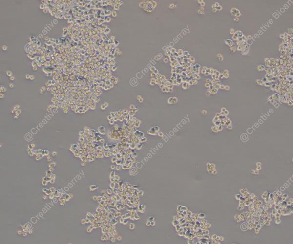
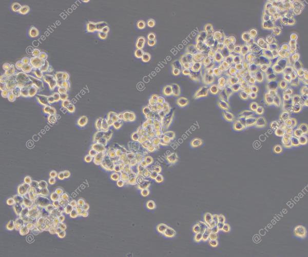
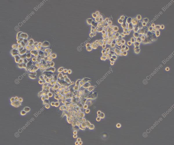
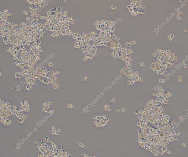
Morphology: other
Culture Properties: Adherent cells
- Specification
- Background
- Scientific Data
- Q & A
- Customer Review
Store in liquid nitrogen.
The GSU cell line is a human cell line derived from gastric cancer, specifically established from the cells present in the ascitic fluid of a patient with this malignancy. Ascites can contain cancer cells that have detached from the primary tumor, providing a rich source of malignant cells for cell line development. The GSU cell line has been characterized by its growth properties, genetic profile, and phenotypic traits, making it a valuable tool for cancer research.
The GSU cell line was created to provide a reliable and reproducible model system for studying the biology of gastric cancer. In the laboratory, GSU cells exhibit characteristics typical of gastric cancer cells, such as rapid proliferation, the ability to form colonies in soft agar, and the expression of specific biomarkers associated with gastric cancer. These cells can be cultured in vitro, allowing researchers to perform a variety of experiments to investigate the effects of genetic mutations, drug treatments, and other interventions on gastric cancer cell behavior.
Trastuzumab and Cetuximab Sensitivity of Gastric Cancer Cell Lines
A panel of 11 gastric cancer cell lines was used. Following treatment with 10 µg/ml cetuximab, GSU cells displayed a decrease in cell proliferation to 68.10% in comparison with the untreated control (p = 0.04). H111TC cells were less sensitive with growth rates of 82.30% after treatment with 10 µg/ml cetuximab (p = 0.002) and a maximum inhibition of 80.16% after application of 200 µg/ml (p < 0.001). MKN7 cells showed only a minor sensitivity with a maximum decrease of cell proliferation to 83.63% following treatment with 100 µg/ml cetuximab (p = 0.001). HGC-27 cells were completely resistant to cetuximab.
Additionally, the trastuzumab sensitivity of the cell lines was investigated via cell proliferation assay (Fig. 1). Two cell lines were identified as trastuzumab sensitive: GSU and H111TC. Both displayed a significant decrease in cell proliferation after the application of the therapeutic. For GSU, 0.1 µg/ml was already sufficient to inhibit the proliferation rate to 80.10% (p = 0.009), while 40 µg/ml caused an inhibition of 72.98% (p = 0.002). H111TC displayed a less sensitive phenotype with a maximum inhibition of 80.03 and 80.31% after application of 20 µg/ml and 40 µg/ml trastuzumab (p = 0.024; p = 0.02), respectively. For the other cell lines, no significant inhibition of cell proliferation was observed.
Table 1. Effect of cetuximab treatment on the gastric cancer cell lines. (Kneissl J, et al., 2017)
| Cell lines | AGS | GSU | H111TC | HGC-27 | Hs746T | KATOIII | LMSU | MKN1 | MKN7 | MKN28 | MKN45 |
| Cetuximab sensitivity | - | +++ | ++ | - | - | - | - | ++ | + | ++ | - |
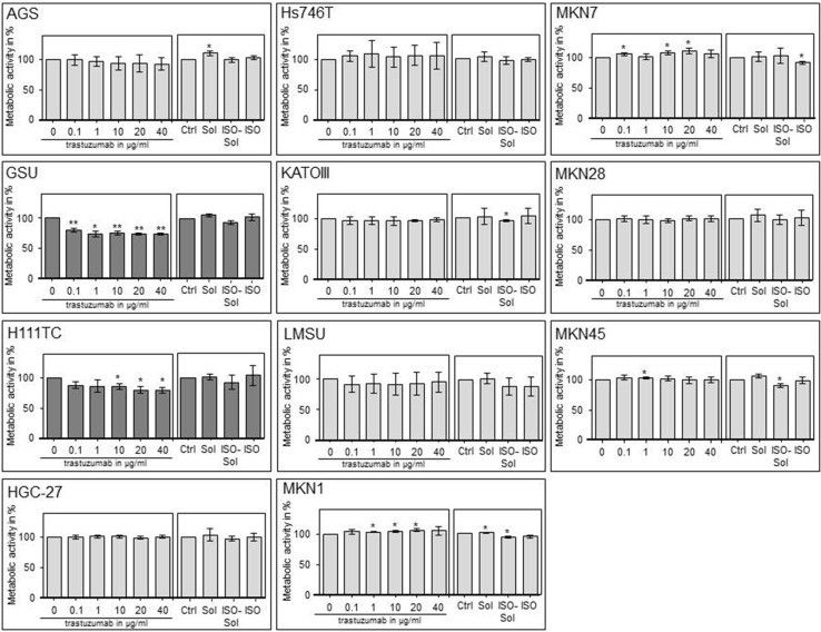 Fig. 1 Effect of trastuzumab treatment on the cell proliferation of gastric cancer cell lines. (Kneissl J, et al., 2017)
Ask a Question
Write your own review
- You May Also Need

Description: Small cell gastrointestinal carcinoma showing creatine kinase-BB and aromatic L-amino-acid decarboxy. Cell growth is slow.

Description: Human cell line derived from signet ring cell carcinoma of stomach.

Description: Human cell line derived from stomach cancer (gastric cancer; highly differentiated tubular adenocarcinoma).

Description: Human gastric cancer cell line derived from metastasis at liver.
- Adipose Tissue-Derived Stem Cells
- Human Neurons
- Mouse Probe
- Whole Chromosome Painting Probes
- Hepatic Cells
- Renal Cells
- In Vitro ADME Kits
- Tissue Microarray
- Tissue Blocks
- Tissue Sections
- FFPE Cell Pellet
- Probe
- Centromere Probes
- Telomere Probes
- Satellite Enumeration Probes
- Subtelomere Specific Probes
- Bacterial Probes
- ISH/FISH Probes
- Exosome Isolation Kit
- Human Adult Stem Cells
- Mouse Stem Cells
- iPSCs
- Mouse Embryonic Stem Cells
- iPSC Differentiation Kits
- Mesenchymal Stem Cells
- Immortalized Human Cells
- Immortalized Murine Cells
- Cell Immortalization Kit
- Adipose Cells
- Cardiac Cells
- Dermal Cells
- Epidermal Cells
- Peripheral Blood Mononuclear Cells
- Umbilical Cord Cells
- Monkey Primary Cells
- Mouse Primary Cells
- Breast Tumor Cells
- Colorectal Tumor Cells
- Esophageal Tumor Cells
- Lung Tumor Cells
- Leukemia/Lymphoma/Myeloma Cells
- Ovarian Tumor Cells
- Pancreatic Tumor Cells
- Mouse Tumor Cells