ECC12
Cat.No.: CSC-C6434J
Species: Homo sapiens (Human)
Source: Stomach
Morphology: epithelial-like
Culture Properties: Adherent cells
- Specification
- Background
- Scientific Data
- Q & A
- Customer Review
Store in liquid nitrogen.
The ECC12 cell line is a model for small-cell gastrointestinal carcinoma, a rare and aggressive subtype of gastrointestinal cancer. Small cell carcinomas are characterized by their small, round, and uniform appearance under the microscope, and they are known for their rapid growth and early metastasis.
Unlike many gastrointestinal cancers, which are typically adenocarcinomas, the ECC12 cell line represents a neuroendocrine tumor of the gastrointestinal tract. These types of tumors arise from the specialized neuroendocrine cells found throughout the digestive system. The expression of creatine kinase-BB and aromatic L-amino-acid decarboxylase, as observed in the ECC12 cell line, are characteristic markers of this neuroendocrine phenotype.
Interestingly, the ECC12 cell line exhibits a relatively slow growth rate compared to other small-cell carcinoma models. This distinction may provide important insights into the underlying biological mechanisms that govern the proliferation and aggressiveness of these rare gastrointestinal tumors.
The development of the ECC12 cell line has been a valuable contribution to the field of gastrointestinal cancer research. As a model system, it allows researchers to investigate the unique molecular and cellular features of small-cell gastrointestinal carcinoma, which is crucial for improving our understanding of this understudied disease.
Validation of GNEC Cell Lines and Drug Sensitivity
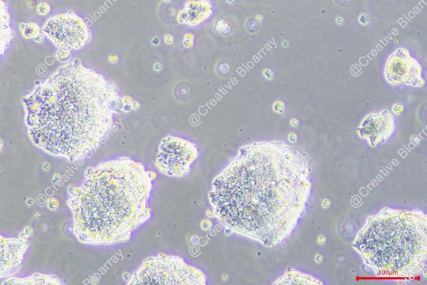
Gastric neuroendocrine carcinoma (GNEC) is a rare cancer detected in the stomach. ECC10 and ECC12 were the only two GNEC cell lines available. Both cell lines are in small cell type and grow in grape-like clusters – "formation of cell clump" (Fig. 1A). To confirm the reliability of these two cell lines, the levels of the two GNEC diagnosis makers CgA and SYN were detected in ECC10 and ECC12. In comparison to GAC cell lines MGC-803 and AGS, the mRNA levels (Fig. 1B) and protein levels (Fig. 1C) of CgA and SYN were significantly higher in ECC10 and ECC12, assuring the validity of these two cell lines for further study. Firstly, the fold change of the top 6 DEGs from the above RNA-seq analyses was selected for expression detection in two GNEC and seven GAC cell lines. The expression patterns of these targets were correlated with the transcriptome results of GNEC and GAC tumor samples (Fig. 1D-I). Next, the RNA levels of 42 DEGs (26 up-regulated genes and 16 down-regulated genes) with the most significant fold changes which were listed after the above 6 targets had been chosen for further validation in GNEC and GAC cell lines. The GNEC cell lines did demonstrate a similar unique gene expression pattern as GNEC patient tissue samples, further confirming the validity of these two GNEC cell lines. Then in GAC (AGS and MGC-803) and GNEC (ECC10 and ECC12) cell lines, the efficacy of chemotherapy drugs commonly waa tested used for GNEC patients including 5-fluorouracil, oxaliplatin, cisplatin, and irinotecan. The two GNEC cell lines displayed lower sensitivity to chemotherapy drugs than the two GAC cell lines (Fig. 1J, K, L, and M).
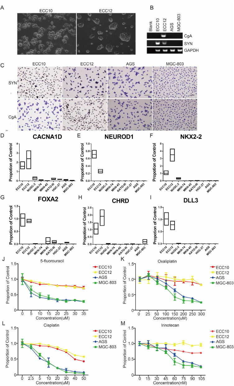 Fig. 1 (A) ECC10 and ECC12 live cell image. (B) CgA and SYN transcriptional level detection in ECC10 and ECC12 by PCR. (C) CgA and SYN transcriptional level detection in ECC10 and ECC12 by IHC. (D-I) Validation of RNA-Seq data by quantitative PCR showed 6 selected genes in fold change top 48. (J-K) Common chemotherapy agents were tested in GNEC and GAC cell lines. Compared to GAC cell lines AGS and MGC-803, GNEC cell lines ECC10 and ECC12 have lower sensitivity to 5-fluorouracil, oxaliplatin, cisplatin, and irinotecan treatment. (Xie J, et al., 2020)
CDK5RAP3 in GNEC Cells Indirectly Affects Vein Endothelial Cell Migration and Tube Formation in Vitro
Cyclin-dependent kinase 5 regulatory subunit associated protein 3 (CDK5RAP3, or C53 for short) was first identified as a binding protein for CDK5 activator proteins p35 and p39. CDK5RAP3 expression varies significantly in different human tumors. The basic expression of CDK5RAP3 from ECC10 and ECC12 was detected by Western blot. The expression of CDK5RAP3 in ECC10 cells was lower than in ECC12 cells. To examine the effect of CDK5RAP3 on GNEC cells, ECC10 and ECC12 cells were created with stable overexpression or knockdown of CDK5RAP3. Changes in CDK5RAP3 expression were confirmed by both Western blot and RT-PCR (Fig. 2a, b). Because CDK5RAP3 was isolated as a binding protein of the Cdk5 activator p35, a Western blot was performed to detect the expression of CDK5, p35, and p39. The results showed that the expression levels of these proteins showed a trend consistent with CDK5RAP3.
Since angiogenesis involves endothelial cell migration and tube formation, HUVEC recruitment assays and wound healing and tube formation assays were performed to gain insight into the role of CDK5RAP3 in GNEC. In brief, TCM from cells with stable overexpression or knockdown of CDK5RAP3 or control was collected and added to the lower chamber with the HUVECs in the upper chamber of the Transwell system. Compared to the control group, TCM from stably knocked down CDK5RAP3 cells exhibited significantly enhanced HUVEC cell migration (Fig. 2c, d) and tube formation potential (Fig. 2e, f). In contrast, TCM from stably overexpressed CDK5RAP3 cells exhibited decreased migration (Fig. 2c, d) and tube formation abilities (Fig. 2e, f). Similar patterns were observed in wound healing (Fig. 2g, h), suggesting that dysregulation of CDK5RAP3 in GNEC cells may indirectly affect EC migration and tube formation in vitro.
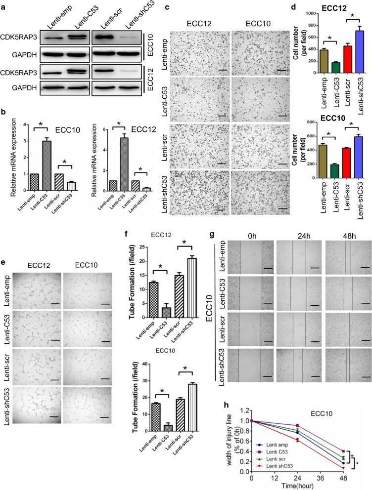 Fig. 2 Endothelial cell migration and tube formation are indirectly influenced by CDK5RAP3 in GNEC cells. ECC10 and ECC12 cells with stable overexpression or knockdown of CDK5RAP3 were created. (Diaz-Lagares A, et al., 2016)
Ask a Question
Write your own review
- You May Also Need

Description: Human cell line derived from signet ring cell carcinoma of stomach.

Description: Human cell line derived from stomach cancer (gastric cancer; highly differentiated tubular adenocarcinoma).

Description: Human gastric cancer cell line derived from metastasis at liver.

Description: Species: human female 55 years old; Tissue: colon; Tumor: adenocarcinoma
- Adipose Tissue-Derived Stem Cells
- Human Neurons
- Mouse Probe
- Whole Chromosome Painting Probes
- Hepatic Cells
- Renal Cells
- In Vitro ADME Kits
- Tissue Microarray
- Tissue Blocks
- Tissue Sections
- FFPE Cell Pellet
- Probe
- Centromere Probes
- Telomere Probes
- Satellite Enumeration Probes
- Subtelomere Specific Probes
- Bacterial Probes
- ISH/FISH Probes
- Exosome Isolation Kit
- Human Adult Stem Cells
- Mouse Stem Cells
- iPSCs
- Mouse Embryonic Stem Cells
- iPSC Differentiation Kits
- Mesenchymal Stem Cells
- Immortalized Human Cells
- Immortalized Murine Cells
- Cell Immortalization Kit
- Adipose Cells
- Cardiac Cells
- Dermal Cells
- Epidermal Cells
- Peripheral Blood Mononuclear Cells
- Umbilical Cord Cells
- Monkey Primary Cells
- Mouse Primary Cells
- Breast Tumor Cells
- Colorectal Tumor Cells
- Esophageal Tumor Cells
- Lung Tumor Cells
- Leukemia/Lymphoma/Myeloma Cells
- Ovarian Tumor Cells
- Pancreatic Tumor Cells
- Mouse Tumor Cells