
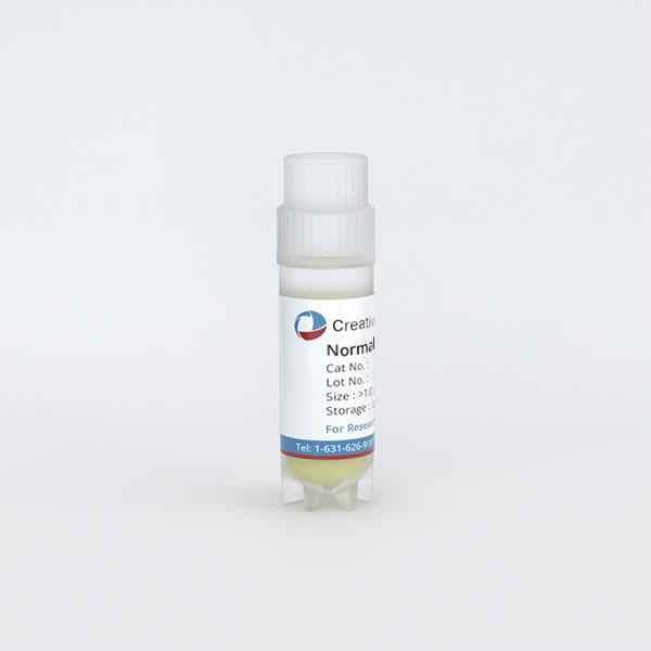
Normal PB Monocyte Derived Macrophages
Cat.No.: CSC-C4427X
Species: Human
Source: Peripheral Blood; Blood
Cell Type: Macrophage
- Specification
- Background
- Scientific Data
- Q & A
- Customer Review
Normal Human Peripheral Blood Monocyte-Derived Macrophages are primary macrophages generated through the in vitro differentiation of circulating monocytes obtained from healthy donors. They provide a human cellular model for investigating innate immune responses, phagocytosis, and inflammatory regulation.
These cells enable investigation of macrophage responses in a nonmalignant cellular background and provide a reference for comparisons with disease-associated populations. Studies comparing primary monocyte-derived macrophages with THP-1-derived models have identified differences in polarization and phagocytic behavior, highlighting the importance of primary cells when evaluating human macrophage function.
Their functional versatility supports complementary assays of microbial particle uptake, apoptotic-cell clearance, cytokine secretion, and cellular metabolism. Fc-dependent phagocytosis also enables investigation of antibody-mediated target uptake. These measurable responses make monocyte-derived macrophages useful for mechanistic studies and evaluation of compounds that influence inflammatory signaling or phagocytic activity.
Another strength is their responsiveness to defined environmental signals. Differentiation factors such as M-CSF or GM-CSF, followed by selected activation stimuli, allow researchers to investigate distinct macrophage programs under controlled conditions.
Soft Tissue-Mimicking Hydrogel Stiffness Modulates Polarization of Human Monocyte-Derived Macrophages
Minimizing the foreign body response (FBR) to implants remains an unmet clinical challenge. The mechanical properties of biomaterials, particularly stiffness, have been shown to influence macrophage activation and FBR. However, current literature on the effects of material stiffness on macrophage activation presents conflicting results. These inconsistencies may stem from the use of cell lines or murine primary cells, which do not fully represent the behavior of primary human monocyte-derived macrophages. Additionally, many previous studies have focused on stiffness values that fall outside the physiological range of soft tissues.
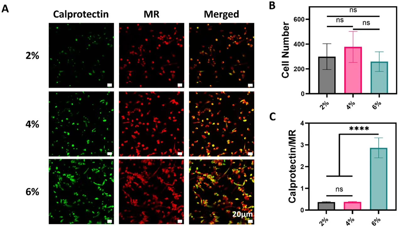
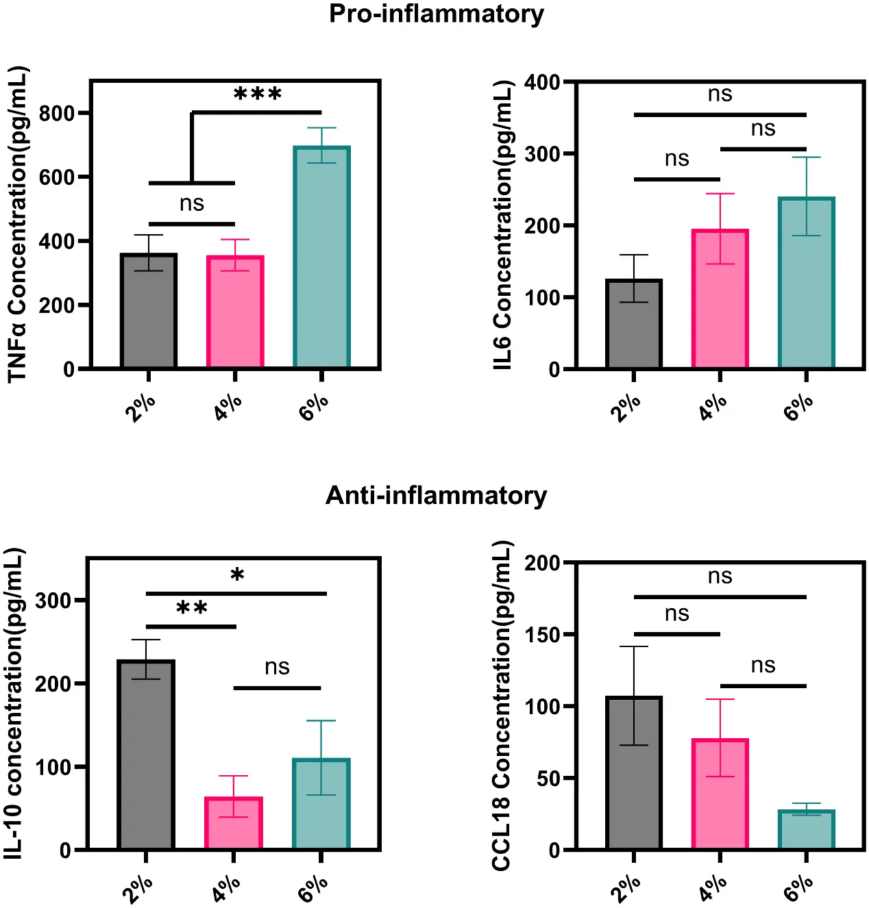
In this study, Coser, Consuelo investigated how variations in stiffness affect the activation status of human monocyte-derived macrophages using alginate methacrylate (ALMA) hydrogels. The hydrogel stiffness was tuned within a physiologically relevant range (0.25–4.5 kPa) to mimic the mechanical properties of soft tissues. They found that increasing hydrogel stiffness consistently upregulated pro-inflammatory markers. Specifically, the stiffest hydrogel (ALMA 6% w/v) induced higher secretion of TNF-α and increased the calprotectin-to-mannose surface maker ratio, both hallmarks of inflammatory macrophages. Moreover, macrophages cultured on stiffer hydrogels exhibited a more elongated morphology and greater spreading. These findings provide new insight into how small changes in stiffness, within a soft tissue-relevant range, can modulate the inflammatory behavior of human macrophages.
Ask a Question
Write your own review
Description: Chronic Myeloid Leukemia-PB Plasma is obtained by centrifuging Chronic Myeloid Leukemia peripheral blood. Plasma has a diminished number of cells and platelets. Chronic Myeloid Leukemia plasma is ...
Description: Acute Myeloid Leukemia-PB-Plasma is obtained by centrifugation of Acute Myeloid Leukemia peripheral blood. Acute Myeloid Leukemia-PB-plasma has a diminished number of cells and platelets. Acute ...

Description: These mononuclear cells (Mononuclear Cells) are collected using the COBE Spectra apheresis machine (>90% of the cells are Mononuclear Cells). These MPB-Mononuclear Cells products have been depleted ...
Description: NPB-Macrophages are derived from negatively-selected peripheral blood monocytes and stimulated with M-CSF and IL-4 growth factors and cultured for 6-7 day.
Description: PV-PB-Mononuclear Cells are enriched by separation using Ficoll-Paque density gradient from the peripheral blood. PV-PB-Mononuclear Cells are available in untreated and relapsed/refractory stages.

Description: Acute Lymphoid Leukemia-PB-Plasma is obtained by centrifugation of Acute Lymphoid Leukemia peripheral blood. This product has a diminished number of cells. Acute Lymphoid Leukemia-Plasma are ...