JC-1 Mitochondrial Membrane Potential Assay
Introduction
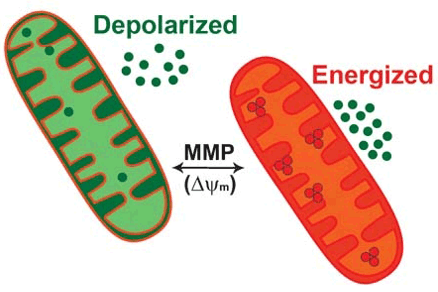
JC-1 mitochondrial membrane potential assay is a fluorescence-based method to track the behavior of mitochondria. Mitochondrial membrane potential (MMP) is an important parameter of mitochondrial function that has been used as an indicator of cell apoptosis. Several cell membrane permeable fluorescent dyes, such as rhodamine-123 (Rh-123), 3, 3′-dihexyloxacarbocyanine iodide (DiOC6), 5,5’,6,6’-tetrachloro-1,1’,3,3’-tetraethylbenzimi-dazolylcarbocyanine iodide (JC-1), tetramethyl rhodamine methyl and ethyl esters (TMRM and TMRE), are currently available to measure changes in MMP. JC-1 is a cationic dye that accumulates in energized mitochondria. In apoptotic cells with low MMP, JC-1 remains in a monomeric form, which exhibits green fluorescence. While in healthy cells with high MMP concentrations, JC-1 forms complexes known as J-aggregates with intense red fluorescence. The higher the ratio of red to green fluorescence, the higher the polarization of the mitochondrial membrane.
JC-1 Staining Solution Preparation
Thaw all the JC-1 reagent at room temperature before use. Prepare a staining solution by diluting the reagent according to the instructions. Dissolve and mix well to make sure there are no particles or flakes in the solution.
The JC-1 staining solution is required to be 1 mL/well of the 6-well plate, and other culture vessels is deduced by analogy, for cell suspension, 0.5 mL of JC-1 staining working solution is required for every 0.5-1 million cells.
Assay Procedure
- Culture and treat cells according to your experimental protocol to induce apoptosis.
For negative control, treat cells with vehicle only.
For positive control, treat cells with FCCP or CCCP at 5-50 μM for 15 to 30 minutes. Typically, the membrane potential of mitochondria is completely lost after 15 minutes of treatment with 50μM CCCP. The concentration and duration of CCCP may vary for a particular cell. It is necessary to refer to the relevant literature to determine and optimize the condition. - Add JC-1 staining solution to each well of the plate and mix gently.
- Incubate the samples for 15-30 minutes. Sufficient staining is usually obtained after 15 minutes. The appropriate incubation time depends on the cell types and cell concentrations, please optimize the incubation time for each experiment.
- Centrifuge the plate for 5 minutes at 400 x g and discard the supernatant carefully.
- Add assay buffer to each well respectively.
- Centrifuge the plate at 400 x g for 5 minutes. Carefully aspirate the supernatant.
- Monitor the fluorescence intensity. Negative controls with mainly JC-1 J-aggregates can be detected by using an excitation of 540 nm and an emission at 590 nm (Ex/Em = 540/590 nm). Apoptotic or unhealthy cells with mainly JC-1 monomers can be detected at excitation and emission wavelengths of 485 and 535 nm (Ex/Em = 485/535 nm).
Notes
- A 6-, 12-, 24-, or 96-well culture plate can be used for this method. It is recommended that the cell be no more than 80% confluent by the time of visualization.
- JC-1 staining solution is difficult to prepare in aqueous medium due to its low solubility and tendency to form particulates that are difficult to eliminate, so make sure JC-1 reagent is completely thawed before diluting it into culture medium.
- All staining procedures must be performed without direct exposure to intense light, as JC-1 is light sensitive.
- If staining is too intense, bring down the final JC-1 concentration may be required.
- It is imperative that samples be analyzed immediately following completion of the staining.
- Buffer or media must be present in the wells during reading of the signal. Don’t allow wells to dry out.
References
- Srilatha S, et al.; Mitochondrial membrane potential assay. Methods Mol Biol, 2016, 1473: 17-22.
- Petit P. X.; et al.; Alterations in mitochondrial structure and function are early events of dexamethasone-induced thymocyte apoptosis. J. Cell Biol. 1995, 130 (1): 157-167.
Cell Services:
Cell Line Testing and Assays: