Comparative Research of IHC and FISH in HER 2 / Neu Gene Detection
International Journal of Hematology-Oncology Stem Cell Research. 2022 Oct 1; 16 (4): 217-223.
Authors: Aznab M, Izadi B, Amirian F, Khazaei S, Madani SH, Ramezani M.
INTRODUCTION
Amplification of HER2 is an important factor in the diagnosis and treatment of breast cancer. Fluorescence in situ hybridization (FISH) is the gold standard for the detection of HER2-positive tumors. However, the Immunohistochemistry (IHC) assay for the detection of HER2 is more popular in the preclinical laboratory since it is faster and more economical compared to the FISH test.
METHODS
- Patient Samples. In this descriptive-analytical study, 44 paraffin blocks with breast carcinoma were collected from the specimens of patients admitted to the pathology laboratory of Imam Reza Hospital in Kermanshah.
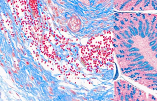
- Hematoxylin and Eosin (H and E) Staining. Paraffin-embedded tissues were stained with hematoxylin and eosin (H and E) method as 4 μm sections. In this technique, glass slides containing sections of tissue were incubated for 2 hours at 70°C. Slides were then soaked in several containers filled with xylene, the graded sequence of ethanol solutions, hematoxylin, lithium carbonate, and eosin.
- Immunohistochemistry. Immunohistology staining has been performed on formalin-fixed paraffin-sealed tissue sections using Her2 antibodies. 4 μm sections of tissue were deparaffinized in a hot air oven for 24 hours at 60-65°C. Slides were immersed in a Tris buffer jar (pH = 9) and warmed for 20 minutes in the autoclave at 121°C followed by washing in PBS solution to retrieve antigens. the slides were incubated with primary and secondary antibodies at 45-120 and 30-45 minutes, respectively in a humid and dark place at room temperature. The slides were coated with a chromogenic surface solution known as 3,3'-diaminobenzidine (DAB) for 5 minutes. The counterstaining was performed for 30-60 seconds with hematoxylin and washed in water. Stained slides were immersed in graded ethanol series and then xylene for transparency and dehydration of tissues. Afterward, slides were mounted under a microscope for examination.
- Browse our recommendations
Creative Bioarray offers a range of high-quality products and services for our customers' research, including but not limited to the products in the table below.
| Product/Service Types | Description | Recommended Products |
| Hematoxylin and Eosin (H and E) Staining | HE staining is a common staining method used in histology. This staining method is based on the fact that tissue structures have different levels of binding to different dyes. | Hematoxylin staining solution, Eosin staining solution,… |
| Immunohistochemistry | IHC is a routine method to detect antigens in cells of a tissue section. It is widely used in the diagnosis of diseases including cancer, neurological disease, digestive disease, etc. | Immunohistochemistry (IHC), Immunofluorescence (IF) Service, Tris Buffer pH 10.1, Steady DAB/Plus, Xylene substitute solution… |
| FISH | FISH is a powerful and easy method to detect RNA or DNA sequences in cells, tissues, and tumors. | Fluorescent In Situ Hybridization (FISH) Service, FISH Probe Design, Synthesis and Testing Service, FFPE FISH Pretreatment Kit… |
RESULTS
- Forty-four patients were included in the study. In the FISH technique, HER2 + was detected in 21 (47.7%) out of 44 patients, and HER2- was detected in 23 (52.3%) patients. Similarly, in the IHC technique, 3 (6.8%) patients had IHC 3 + (positive), 5 (11.4%) had IHC 0 and IHC +1 (Negative), and in 36 (81.8%) patients, IHC 2+ (Equivocal) was statistically significantly different from standard FISH method (P = 0.019).
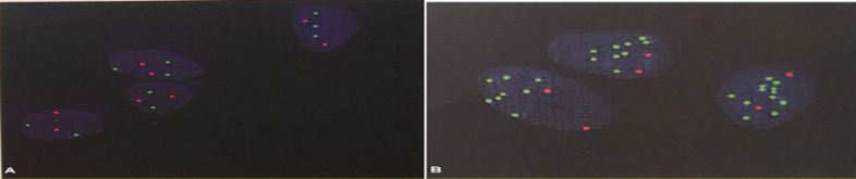 Fig. 1 FISH images of an HER 2gene amplified sample using HER 2probes and centromere chromosome 17 (red).
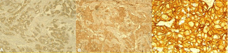 Fig. 2 IHC images of a HER2 gene amplified sample using anti-HER2 antibodies.
- The HER2 gene is significantly more amplified in patients who entered menopause than in other women (P = 0. 035), and there was no statistically significant association between HER2 gene proliferation and estrogen, progesterone receptors, or the P53 and KI67 genes.
SUMMARY
This result demonstrated that the IHC test is not a reliable test to determine HER2 amplification. This study represented that FISH analysis is more reliable than IHC and must be preferentially performed for all cases, especially for HER2 +2 cases for whom the IHC result is 2+.
RELATED PRODUCTS & SERVICES
Reference
- Aznab M, et al. (2022). "Comparison of Immunohistochemical Methods (IHC) and Fluorescent in Situ Hybridization (FISH) in the Detection of HER 2 /Neu Gene in Kurdish Patients with Breast Cancer in Western Iran." Int J Hematol Oncol Stem Cell Res. 16 (4), 217-223.