Cell Spreading Assay
Cell adhesion is a complex process involving many different molecular interactions, including receptor-ligand binding, changes in the fluxes through intracellular signaling pathways, and modulation of cytoskeletal assembly. The ability to quantify adhesion has proven to be extremely valuable for those researchers investigating the molecular mechanisms underlying these processes. The spreading assay, which employs a phase contrast microscopy to measure the flattening of adherent cells, takes longer to perform, but is unlikely to be nonspecific. In addition, by observing the response of the cells in a spreading assay, information on the manner in which the cells react to the substrate can also be obtained. A further advantage of spreading assay is that it is more sensitive when used to measure inhibitory activity because the readout is more reliant than attachment assay on multiple adhesive interactions. Finally, spreading assay does not require replicate wells, it is more economical in their use of substrates.
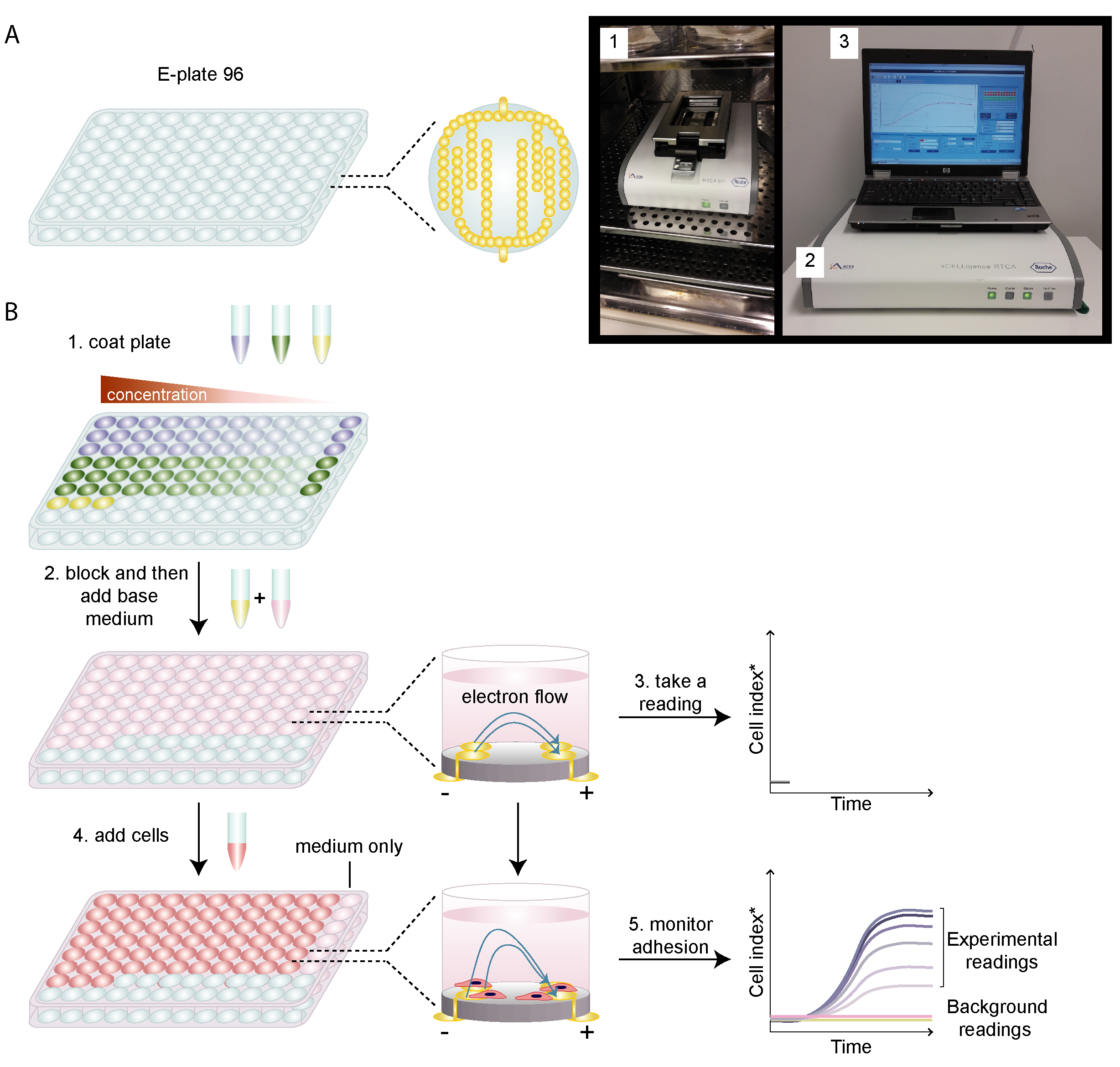 Figure 1. Schematic diagram of cell adhesion events.
Materials
- Bovine serum albumin (BSA) 10 mg/mL
- Collagen I
- Fetal bovine serum (FBS)
- 96-well tissue culture plate
- Dulbecco's modified eagle medium (DMEM)
- 0.5 M EDTA solution (pH 8.0)
- 0.05% (w/v) sodium azide
- 5% (w/v) glutaraldehyde
- Divalent cation-free phosphate buffered saline (PBS)
Assay Procedure
- Grow the cells in DMEM medium supplemented with 10% FBS.
- Prepare 40 μg/mL collagen I solution in PBS, store at 4°C, and prepare 0.1% BSA solution in DMEM.
- Coat the 96-well plate with the collagen I solution at 4°C.
Note: There is no need to carry out spreading assay with replicate wells, because quantitation is performed by counting multiple fields from within the same well. - After 12 h of coating, remove the collagen I solution and air-dry the plate at room temperature in the tissue culture hood.
- Deprive cells of serum for 8 h before the adhesion assay.
- Use 10 mM EDTA in DMEM to detach the cells and then observe them under a microscope to confirm complete dissociation of the cells.
- Wash cells twice with DMEM to remove EDTA, resuspend cells at 2 x 10⁵ cells/mL in DMEM with 0.1% BSA.
- For cell-substratum adhesion assay, add 100 μL cell suspension to each of the collagen-I-coated well. Incubate the plate at 37°C for 20 min to allow the cells to adhere to the surface.
- Add 100 μL DMEM to each well to wash off any non-adherent cells, wash four times.
Note: To achieve consistency, always add/remove DMEM gently with multi-channel pipetter for multiple wells. - After washing, add DMEM with 10% FBS and incubate the cells at 37°C for 4 h for recovery.
- Fix cells by direct addtion of 10 μL 50% (w/v) glutaraldehyde and leave at room temperature for 30 min.
- Aspirate fixative and store cells in PBS (without divalent cations), 0.05% NaN3.
- Determine the percentage of cells that adopt a spread morphology using an inverted phase contrast microscope.
Note: Understandably, the optical quality of the plastic that is used to make microtiter plates is not ideal for phase contrast microscopy. However, the observation of adherent cells can be greatly improved by adding sufficient PBS/azide to form an inverted meniscus at the top of the well and then carefully placing a glass cover slip over the plate.
References
- Amelia A K. et al.; A review of cell adhesion studies for biomedical and biological applications. International Journal of Molecular Science, 2015, 16: 18149-18184.
- Humphries M J. Cell adhesion assays. Methods Mol Biol, 2009, 522: 203-210.
Cell Services:
Cell Line Testing and Assays