ATP Cell Viability Assay
ATP is involved in a variety of enzymatic reactions to maintain normal life activities. When normal cells undergo apoptosis and necrosis, the content of ATP will be characteristic changed. Thus, ATP has been widely accepted as a valid marker of viable cells. The measurement of ATP using firefly luciferase is the most commonly applied method for estimating the number of viable cells.
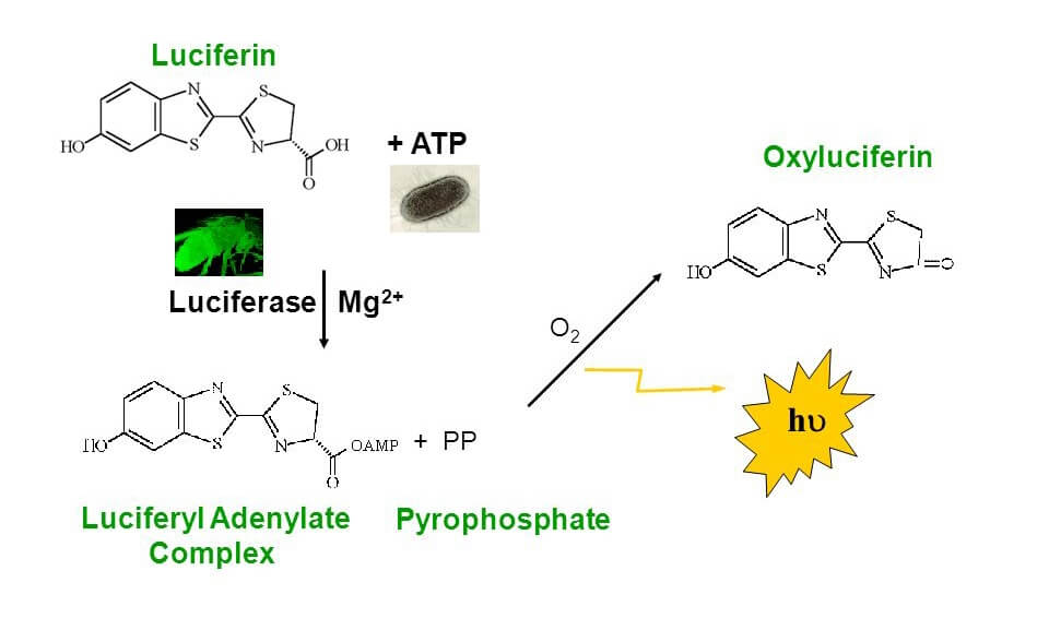
The ATP cell viability assay utilizes luciferase to catalyze the formation of light from ATP and D-luciferin, which can be measured with a luminometer or Beta Counter. The advantage of ATP assay is that you do not have to rely on an incubation step with a population of viable cells to convert a substrate (such as tetrazolium and resazurin) into a colored compound. There is also no need to remove cell culture medium or wash cells before adding the reagent, which can be fully automatic for high throughput.
 Figure 1. The principle of ATP Bioluminescence.
Materials
- Cell Lysate Reagent
- D-Luciferin
- Firefly Luciferase
- ATP
- Assay Buffer
- Microcentrifuge
- Pipettes and pipette tips
- Luminometer plate or Beta Counter
- 96-well plate
- Cuvettes
- Orbital shaker
Standard Curve
For quantifying the amount of ATP amount, a series of ATP standards can be prepared in 100 μL of the same diluent as the samples (Table 1).
Table 1. Preparation of ATP standards by serial dilution in dH2O, PBS, or cell culture medium.
| Volume of ATP solution | Volume of diluent | Final ATP concentration | ATP/100 μL | |
| A | 2.5 μL ATP standard (2 mM) | 500 μL | 10 μM | 1000 pmol |
| B | 50 μL solution A | 450 μL | 1 μM | 100 pmol |
| C | 50 μL solution B | 450 μL | 100 nM | 10 pmol |
| D | 50 μL solution C | 450 μL | 10 nM | 1 pmol |
| E | 50 μL solution D | 450 μL | 1 nM | 0.1 pmol |
| F | -- | 500 μL | 0 | 0 pmol |
| * Transfer 100 μL of each ATP standard to a fresh tube for assay. |
Preparation of ATP Detection Cocktail
- Thaw ATP Assay Buffer and pipette a desired volume (2.5 mL or 25 mL) into a new container.
- In a clean container, dissolve the supplied D-Luciferin with the above Assay Buffer to a final concentration of 0.4 mg/mL.
Note: If you need less than 2.5 mL or 25 mL ATP assay solution as described in step 2, you may prepare a D-Luciferin stock solution in dH2O and store it at -20°C or below for repeated use. The D-Luciferin stock solution should be stable for at least one month. - Add Firefly Luciferase to the ATP assay solution in a ratio of 1 μL to 100 μL (25 μL Luciferase for 2.5 mL or 250 μL Luciferase for 25 mL of the ATP assay solution). ATP Detection Cocktail should be prepared fresh before each use for maximum activity.
Note: Because of the high sensitivity of the ATP assay, avoid contamination with ATP from exogenous biological sources, such as bacteria or fingerprints.
Assay Protocol
- For suspension cells, transfer 10 µL of the cultured cells (containing 103-104 cells) into luminometer plate. Add 100 µL of the Nuclear Releasing Reagent.
- For adherent cells, remove culture medium and treat cells (103-104) with 100 µL of Nuclear Releasing Reagent for 5 minutes at room temperature with gentle shaking.
- Add 1µL ATP Detection Cocktail into the cell lysate. Read the sample in 1 minute in a luminometer.
- The background luminescence should be subtracted from all readings. The amount of ATP in experimental samples can then be calculated from the standard curve.
Note: The assay can be analyzed using cuvette-based luminometers or Beta Counters. The entire assay can also be done directly in a 96-well plate. It can also be programmed automatically using instrumentation with injectors.
References
- Mueller H. et al.; Comparison of the usefulness of the MTT, ATP, and Calcein assays to predict the potency of cytotoxic agents in various human cancer cell lines. Journal of Biomolecular screening, 2004, 9 (6): 506-515.
- Riss T. L. et al.; Cell viability assays. Assay Guidance Manual, 2013.
Cell Services:
Cell Line Testing and Assays: