Protocols of Primary Cultures for IHC-Viability Assays
GUIDELINE
Primary cultures for IHC-viability assays refer to the use of primary cells derived directly from animal or human tissue samples for immunohistochemistry (IHC) analysis aimed at assessing cell viability. These assays are commonly used in research and pharmaceutical applications to evaluate the effects of different compounds or treatments on cell survival and function. The following protocol provides a method of primary cultures for IHC-viability assays.
METHODS
- The mesencephalic neurons and glia are dissociated from neuronal tissue with trypsin (final concentration, 26 mg/ml in 0.9% [w/v] NaCl). Cells are plated on coverslips previously treated with poly-L-lysine (5 mg/ml).
- The media consists of DMEM, 10% (v/v) FBS, 10% (v/v) HS, penicillin (100 U/ml), and streptomycin (100 mg/ml).
- Four days later, the cells were treated for 48 hr with AraC (20 mM) to inhibit the growth of glial cells.
- AraC-treated primary cultures are transduced with lentiviral particles in the presence of polybrene (6 mg/ml). Control cells were incubated without lentivirus.
- After a 72-hour transduction period, the cells were treated with fresh media for an additional 48 hr and analyzed immunocytochemically.
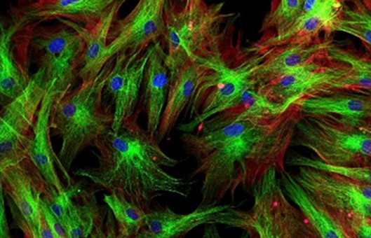
- Primary cells were fixed in 4% (w/v) paraformaldehyde in PBS for 30 min. The cells were permeabilized and blocked simultaneously for 1 hr with PBS containing 1% (w/v) BSA, 10% (v/v) FBS, and 0.3% (v/v) Triton X-100.
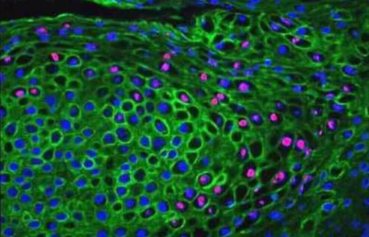
- After washing with PBS, the cells were treated overnight at 4°C with an anti-MAP2 monoclonal IgG (1:500) and an anti-TH polyclonal antibody (1:500) to monitor relative dopaminergic cell viability.
- To determine the lentiviral transduction efficiency, un-transduced cells or cells transduced with lacZ lentivirus (expressing beta-galactosidase fused to the V5 epitope) are treated at room temperature for 1 hr with an anti-V5 monoclonal antibody (1:200) and an anti-MAP2 polyclonal antibody (1:500), or an anti-V5 monoclonal antibody (1:200) and an anti-TH polyclonal antibody (1:500).
- After washing, the cells were treated for 1 hr at room temperature with goat anti-mouse IgG and goat anti-rabbit IgG.
- The primary and secondary antibodies were prepared by diluting stock antibody solutions in PBS with 1% (w/v) BSA.
- The coverslips were mounted onto slides, dried at room temperature overnight, and sealed with clear nail polish.
- MAP2- and TH-immunoreactive primary neurons can be counted in 10 randomly chosen observation fields for each experimental condition at a total magnification of 200x.
- The data should be expressed as the percentage of MAP2-positive neurons that are also TH-positive. Each experiment should be repeated 3 to 4 times using embryonic neurons isolated from independent animals.
Creative Bioarray Relevant Recommendations
- Creative Bioarray offers a comprehensive IHC service from project design, and marker selection to image completion and data analysis. We are dedicated to satisfying every customer and assisting them to achieve their specific research goals.
- We are capable of providing various options for cell viability assays and compound profiling services based on cell viability MTT/MST/WST-8/WST-1/CKK-8 assays, trypan blue assays, calcein-AM assays, fluorescent reactive dye assays, single/two color viability assays, and microplate cell viability assays.
- We also provide our clients with a large and unique collection of high-purity, low-passage human and animal primary cells. Our primary cells cover a wide range of cell types (epithelial cells, endothelial cells, smooth muscle cells, and many other key types) from a large variety of tissues (skin, liver, bone, brain, and more) in both cryopreserved and proliferating formats. Additional isolates (which may be paired, age, gender, haplotype, and/or ethnic-specific) of certain cell types may be available, please contact customer service.
| Product Types | Description |
| Human Primary Cells | Creative Bioarray offers more than 1000 cell types together with selective cell culture media. |
| Animal Primary Cells | Creative Bioarray's animal primary cells are isolated from normal or diseased animal tissues. |
| Primary Cell Media | We provide ready-to-use liquid formulations, as well as custom options for powder media and formula modifications. |
View our full range of human and animal primary cells and find what you need!
NOTES
- Maintain the primary cells in appropriate culture conditions, including the use of suitable growth media, supplements, and growth factors tailored to the specific cell type to ensure cell viability and function.
- Choose an appropriate viability assay based on the specific requirements of the experiment and the characteristics of the primary cells. Common viability assays include MTT assay, MTS assay, FDA/PI staining, or live/dead cell assays using fluorescent dyes.
- For IHC viability assays, the method of fixation and permeabilization of the cells is crucial. Consider using suitable fixatives and permeabilization agents that are compatible with both maintaining cell viability and enabling antibody penetration for detection.
- Select primary and secondary antibodies for the IHC detection of cell viability markers (e.g., cleaved caspases, Annexin V, TUNEL assay) based on their specificity and validation for use in primary cultures. Ensure that the antibodies are suitable for detecting antigens in the relevant cell type and under the chosen culture conditions.