GUIDELINE
- The generation of neurons or glia that can be used to replace lost or damaged tissue is a key step in stem cell-based therapies designed to treat neurodegenerative disease and neurological disorders.
- Several different candidate sources have been proposed for producing these cells. Based upon their proliferative and pluripotential properties, embryonic stem cells (ESCs) are an attractive alternative to fetal or adult-derived central nervous system (CNS) tissue. The derivation of specific neuronal or glial cell types from ESCs invariably includes the production of neural stem cells (NSCs), the self-renewing, pluripotential stem cells of the nervous system, capable of differentiating into neurons, astrocytes, and oligodendrocytes.
METHODS
- The 35 mm Petri dishes are coated with 0.1% gelatin 1 h in advance, and the gelatin is discarded after 1 h of incubation.
- The pluripotent stem cells cultured on the feeder layer were digested with 0.05% Trypsin for 2 min, and the digestion was terminated by adding an equal volume of cell basal medium. Centrifugation is performed at 1000 rpm for 5 min, the supernatant is discarded, and the cells are resuspended with a neural stem cell culture medium. Then, the cells are added to the gelatin-coated 35 mm dishes at a cell density of 2×10⁵ cells per dish, and the cells are left to stand for 30 min at 37°C in a 5% CO2 incubator. The supernatant is aspirated to remove the trophoblastic layer attached to the gelatin.
- The supernatant is centrifuged at 1000 rpm for 5 min, the supernatant is discarded and the cells are resuspended with EB medium, and the cells are inoculated into 100 mm dishes at a density of 1×10⁶ cells per dish, and then incubated at 37°C with 5% CO2 for 48 h. In the following, 3 mL of EB medium was added to each 100 mm dish every day.
- On day 5 of differentiation, 500 μL of 20 μg/mL fibronectin is added to each well of a 12-well plate and left at room temperature overnight.
- On day 6 of differentiation, the fibronectin in the 12-well plate is discarded and 1 mL of EB medium is added to each well. Pipette EB spheres from 100 mm Petri dishes into 12-well plates, and place about 20 EB spheres per well.
- On the 2nd day after EB inoculation for wall apposition, EBM is discarded and 2 mL of N2B27 culture medium is added to each well. The fresh N2B27 culture medium is replaced half daily thereafter.
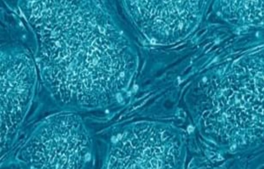
- On day 6 after EB inoculation, a large number of nerve cells and occasionally neurons can be seen crawling out of the wall-adherent balls at this time.
- The cells are digested with 0.05% Trypsin for 2 min and terminated with an equal volume of 10% FBS. 1000 rpm centrifugation is performed for 5 min, and the cells are resuspended with NSCM planted in 35 mm dishes at a density of 5×10⁵ cells per dish, and cultured at 37°C in a 5% CO2 incubator.
- When the cell density reaches 80%, the cells are passaged, usually once in 3-4 d. After several passages, the neural stem cells grew as floating neuro-spheres.
Creative Bioarray Relevant Recommendations
- Creative Bioarray provides embryonic stem cells from both humans and mice. Embryonic stem cells (ES cells) are pluripotent stem cells derived from the inner cell mass of a blastocyst, an early-stage preimplantation embryo.
- We also provide our customers with a large and unique collection of high-purity, low-passage human and animal neural stem cells, including but not limited to Strain C57BL/6 Mouse Neural Stem Cells, Sprague-Dawley (SD) Rat Neural Stem Cells, Human Neural Stem Cells-cortex region, Human iPSC-Derived Neural Stem Cells, Human Neural Stem Cells-ventral mesencephalon region, Mouse Spinal Cord Neural Stem Cells, Rat Hippocampal Neural Stem Cells, and others.
NOTES
- After inoculation of the petri dish, the cell suspension should be shaken well before putting it into the incubator. Otherwise, the EBs tend to stick together.
- Aspirate and add liquid in 12-well plates gently, with the tip of the gun pressed against the wall of the dish to avoid blowing up the EB balls.
RELATED PRODUCTS & SERVICES