
Human Neural Stem Cells-cortex region
- Specification
- Background
- Scientific Data
- Q & A
- Customer Review
Human neural stem cells (NSCs) derived from the cerebral cortex represent a critical experimental system for investigating human corticogenesis, cortical disorders, and regenerative strategies. The cerebral cortex—the seat of higher cognitive functions—contains approximately 16 billion neurons and exhibits extraordinary structural and functional complexity that is uniquely human. Cortical NSCs can be isolated from human fetal telencephalic tissue, typically at 6–9 weeks post-conception, and expanded in vitro as neurospheres or adherent monolayer cultures.
The defining advantages of cortical NSCs lie in three interrelated properties. First, they exhibit robust self-renewal and multipotency, retaining the capacity to generate the three major neural lineages—neurons, astrocytes, and oligodendrocytes—over extended passages. Second, and most critically, they maintain an intrinsic regional identity that is preserved in culture. Upon long-term differentiation in vitro, cortical NSCs generate region-specific neuronal subtypes, including higher proportions of glutamatergic, tyrosine hydroxylase-positive, and calretinin-positive neurons compared to NSCs from other brain regions. This region-specific differentiation potential is retained even after transplantation. Third, when grafted into animal models—including stroke-lesioned rodents—cortical NSCs survive robustly, migrate extensively, and differentiate into mature neurons that integrate into host circuitry, underscoring their strong neurogenic capacity.
These attributes position cortical NSCs as an indispensable platform for modeling human cortical development and neuropsychiatric disorders, screening neuroactive compounds, and developing cell-replacement therapies for cortical pathologies such as stroke and traumatic brain injury. Their ability to recapitulate human-specific corticogenesis in vitro—including the generation of cortical projection neurons and interneurons—offers advantages over rodent models, which lack certain human neural progenitor populations and exhibit different neurogenic trajectories.
Human Neural Stem Cell-Derived Exosomes Protect Against Oxidative Stress-Induced Neuronal Injury In Vitro
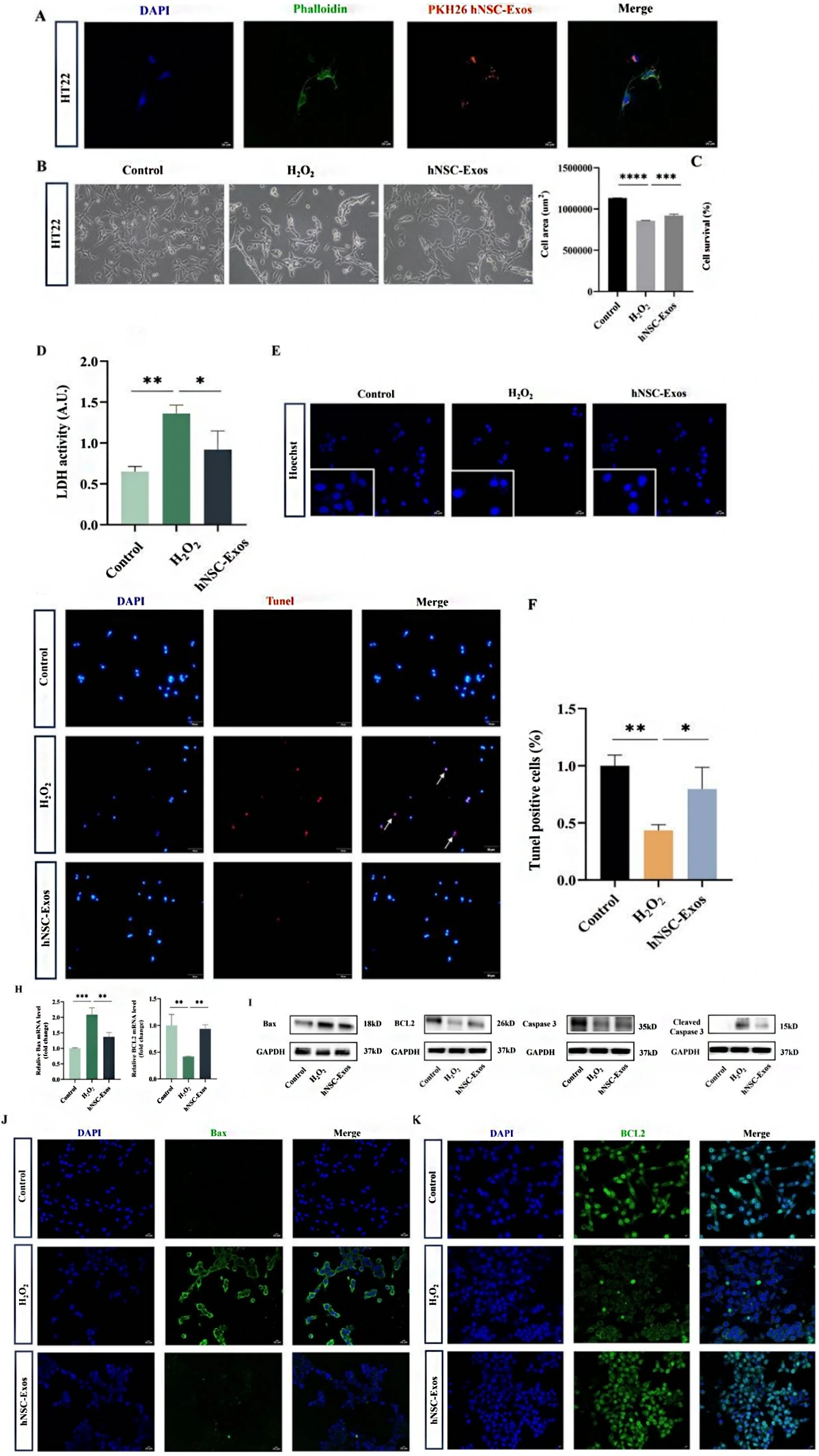
This study investigates the protective effects of hNSC derived exosomes (hNSC-Exos) on neuronal damage. HT22 cells were employed and treated with H2O2 to construct an oxidative stress (OS)-induced injury model. Afterward, hNSC-Exos were co-cultured with neurons, and the internalization process was visualized using fluorescence microscopy.
Successful uptake was evidenced by the presence of PKH26-labeled hNSC-Exos within the cytoplasm of HT22 cells (Fig. 1A). Treatment with hNSC-Exos partially restored the cellular morphology (Fig. 1B), significantly enhanced the percentage of viable cells (Fig. 1C), and mitigated cytotoxicity (Fig. 1D).
Given that H2O2-induced OS is a pathological mechanism implicated in neuronal apoptosis, Hoechst (Fig. 1E), TUNEL (Fig. 1F), annexin V-FITC/PI double staining (Fig. 1G) in conjunction with flow cytometry were employed to assess the protective effects of hNSC-Exos against H2O2-induced apoptosis in HT22 cells. hNSC-Exos markedly diminished the level of H2O2-induced early apoptosis, rather than late apoptosis. qRT-PCR data indicated that the levels of the pro-apoptotic marker Bax were reduced, whereas those of the anti-apoptotic marker BCL2 were elevated following hNSC-Exos treatment (Fig. 1H). WB analysis showed that hNSC-Exos increased the level of Caspase 3 and reduced the level of Cleaved caspase 3 (Fig. 1I), suggesting that hNSC-Exos may have anti-apoptotic or neuroprotective effects. Additionally, immunofluorescence for Bax and BCL2 further corroborated that hNSC-Exos exerted a protective effect against H2O2-induced apoptosis in HT22 cells (Fig. 1J-K).
Ask a Question
Write your own review
- Adipose Tissue-Derived Stem Cells
- Human Neurons
- Mouse Probe
- Whole Chromosome Painting Probes
- Hepatic Cells
- Renal Cells
- In Vitro ADME Kits
- Tissue Microarray
- Tissue Blocks
- Tissue Sections
- FFPE Cell Pellet
- Probe
- Centromere Probes
- Telomere Probes
- Satellite Enumeration Probes
- Subtelomere Specific Probes
- Bacterial Probes
- ISH/FISH Probes
- Exosome Isolation Kit
- Human Adult Stem Cells
- Mouse Stem Cells
- iPSCs
- Mouse Embryonic Stem Cells
- iPSC Differentiation Kits
- Mesenchymal Stem Cells
- Immortalized Human Cells
- Immortalized Murine Cells
- Cell Immortalization Kit
- Adipose Cells
- Cardiac Cells
- Dermal Cells
- Epidermal Cells
- Peripheral Blood Mononuclear Cells
- Umbilical Cord Cells
- Monkey Primary Cells
- Mouse Primary Cells
- Breast Tumor Cells
- Colorectal Tumor Cells
- Esophageal Tumor Cells
- Lung Tumor Cells
- Leukemia/Lymphoma/Myeloma Cells
- Ovarian Tumor Cells
- Pancreatic Tumor Cells
- Mouse Tumor Cells