
Human iPSC-Derived Neural Stem Cells
- Specification
- Background
- Scientific Data
- Q & A
- Customer Review
Never can primary cells be kept at -20 °C.
Human induced pluripotent stem cell-derived neural stem cells (hiPSC-NSCs) are generated through the reprogramming of somatic cells—typically skin fibroblasts or peripheral blood mononuclear cells—back to a pluripotent state, followed by directed neural induction and expansion under defined culture conditions. Functionally resembling embryonic stem cell-derived neural progenitors while circumventing the associated ethical constraints, hiPSC-NSCs have emerged as a transformative platform for neurological research and regenerative medicine.
Their advantages over alternative NSC sources are fourfold. First, patient-specific autologous sourcing – hiPSC-NSCs can be generated from the patient’s own somatic cells, enabling personalized therapies and fundamentally eliminating the risk of immune rejection and the need for lifelong immunosuppression. This autologous paradigm distinguishes them from fetal or allogeneic NSCs.
Second, virtually unlimited scalability – hiPSCs exhibit robust self-renewal, supporting the derivation of continuous, expandable NSC lines amenable to large-scale, clinical-grade manufacturing under current Good Manufacturing Practice (cGMP) standards. This addresses the critical supply limitations inherent to primary fetal tissue.
Third, multidirectional differentiation and regional patterning – hiPSC-NSCs retain multipotency, spontaneously differentiating into neurons, astrocytes, and oligodendrocytes upon mitogen withdrawal. Critically, they can be directed toward region-specific neuronal subtypes—dopaminergic, cortical, or spinal—through patterned induction, offering versatility for modelling distinct neurological conditions.
Fourth, amenability to genetic modification – hiPSC-NSCs are readily compatible with CRISPR-based gene editing, enabling isogenic disease modelling, correction of pathogenic mutations, and ex vivo gene therapy for monogenetic CNS disorders.
Furthermore, long-term transplantation studies have demonstrated robust engraftment, stable differentiation, and no evidence of tumor formation, reinforcing their safety profile. The world's first clinical trial using hiPSC-NSCs for spinal cord injury has already been initiated, underscoring their translational momentum.
Human-Induced Pluripotent Stem Cell–Derived Neural Stem Cell Exosomes Improve Blood–Brain Barrier Function After Intracerebral Hemorrhage
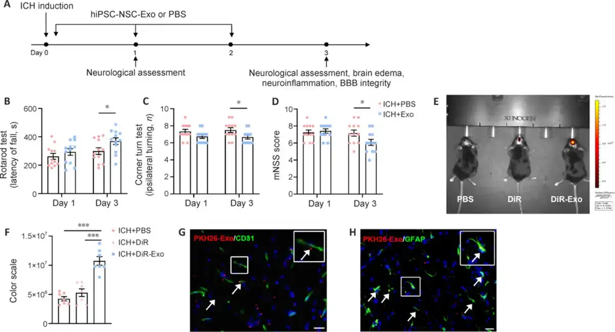
Cerebral edema caused by blood–brain barrier injury after intracerebral hemorrhage (ICH) is an important factor leading to poor prognosis. Human-induced pluripotent stem cell–derived neural stem cell exosomes (hiPSC–NSC–Exos) have shown potential for brain injury repair in central nervous system diseases. To test the effect of hiPSC–NSC–Exos on ICH, a mouse model of ICH was established, and exosomes were administered via intranasal delivery at a dose of 5 μg/g body weight once daily for 3 consecutive days. Neurobehavioral tests were conducted on day 1 and 3 (Fig. 1A).
The performance of the hiPSC–NSC–Exo group in the rotarod and corner turn tests, as well as the modified neurological severity score, were superior to those observed in the control group (Fig. 1B–D). In vivo small animal fluorescence imaging revealed DiR-labeled exosomes clustered near the lesion in the exosome group (Fig. 1E and F). In addition, fluorescence colocalization analysis showed that PKH-26–labeled exosomes were internalized by endothelial cells and astrocytes (Fig. 1G and H). These results suggest that intranasal delivery of hiPSC–NSC–Exos can improve neurological deficits in ICH mice.
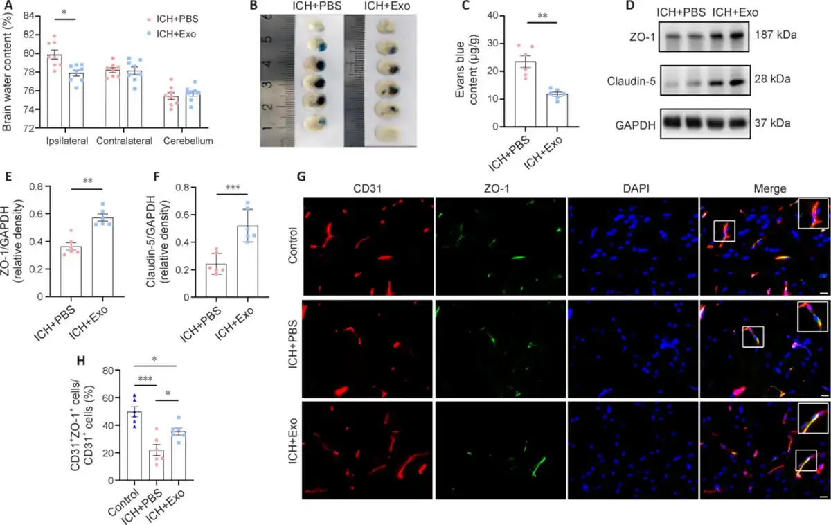
Additionally, hiPSC–NSC–Exo treatment significantly reduced the ipsilateral hemispheric water content (Fig. 2A). EB leakage analysis indicated that hiPSC–NSC–Exos reduced BBB permeability (Fig. 2B and C). To investigate the effects of hiPSC–NSC–Exos on BBB structural components and integrity, the tight junction proteins claudin-5 and ZO-1 were analyzed. Compared with the PBS group, significantly higher levels of claudin-5 and ZO-1 were present around the hemorrhagic lesions in the exosome-treated group (Fig. 2D–F). Double immunofluorescence staining confirmed these findings (Fig. 2G and H).
Ask a Question
Write your own review
- Adipose Tissue-Derived Stem Cells
- Human Neurons
- Mouse Probe
- Whole Chromosome Painting Probes
- Hepatic Cells
- Renal Cells
- In Vitro ADME Kits
- Tissue Microarray
- Tissue Blocks
- Tissue Sections
- FFPE Cell Pellet
- Probe
- Centromere Probes
- Telomere Probes
- Satellite Enumeration Probes
- Subtelomere Specific Probes
- Bacterial Probes
- ISH/FISH Probes
- Exosome Isolation Kit
- Human Adult Stem Cells
- Mouse Stem Cells
- iPSCs
- Mouse Embryonic Stem Cells
- iPSC Differentiation Kits
- Mesenchymal Stem Cells
- Immortalized Human Cells
- Immortalized Murine Cells
- Cell Immortalization Kit
- Adipose Cells
- Cardiac Cells
- Dermal Cells
- Epidermal Cells
- Peripheral Blood Mononuclear Cells
- Umbilical Cord Cells
- Monkey Primary Cells
- Mouse Primary Cells
- Breast Tumor Cells
- Colorectal Tumor Cells
- Esophageal Tumor Cells
- Lung Tumor Cells
- Leukemia/Lymphoma/Myeloma Cells
- Ovarian Tumor Cells
- Pancreatic Tumor Cells
- Mouse Tumor Cells