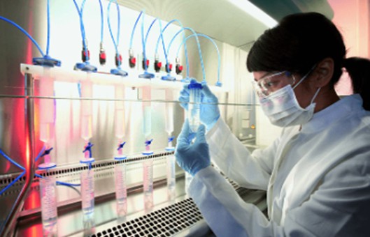
GUIDELINE
-
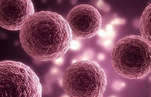
Mouse embryonic fibroblasts are suitable feeder cells for
mouse
or
human embryonic stem cells
and
induced pluripotent stem cells. They can produce factors that inhibit the autonomous differentiation of embryonic stem cells and promote the
proliferation of embryonic stem cells, which can effectively promote the proliferation of cells and maintain
their undifferentiated characteristics and pluripotency.
-
At present, most laboratories use mouse fibroblasts as a feeder layer to isolate and culture embryonic stem
cells. The feeder layer not only provides a stable external environment for the growth and propagation of
embryonic stem cells but also maintains the characteristics of infinite proliferation and differentiation of
embryonic stem cells.
METHODS
Reagents and materials
- Animals. Pregnant mice are prepared for 14 to 16 days of gestation.
-
Sterile anatomical instruments. 2 ophthalmic scissors, 2 ophthalmic forceps, 3 pairs of glass plates, 1 small
pincer, and 1 tissue scissors.
- MEF growth medium. High glucose type DMEM containing 10% fetal bovine serum.
- 35 mm, 60 mm and I00mm tissue culture dishes.
Primary culture
-
Female rats at 12.5 to 14.5 days of gestation are sacrificed with necks broken, and the uterus is exposed under
sterile conditions.
- Lift the proximal cervix with tweezers to separate the mesangium and cut the uterine horn.
-
The whole uterus is removed and placed on a plate with PBS. Wash with PBS three times, and discard the residual
blood on the surface.
-
The uterus is cut open along the mesangial side of the uterus, and the embryo with the fetal membrane is taken
out and placed in another plate containing PBS for full washing, and the surface red blood cells are discarded.
-
Tear the fetal membrane with small tweezers, remove the fetal rat, discard the fetal membrane, and wash with PBS
three times.
-
The head, viscera, and limbs of the embryo are removed, and the trunk is placed in another plate containing PBS.
Wash with PBS at least three times, and the red blood cells are fully discarded.
-
The residual embryo is broken up with ophthalmic scissors, and the paste tissue is transferred to a 20 mL
flat-scale test tube. And 5 mL of trypsin solution and 40 μL of DNase crude product are added to avoid the
release of DNA that would cause the solution to be too sticky.
-
Incubate at 37°C water bath for 30 minutes, or place overnight at 4℃, shake, and then pour the upper cell
suspension into a 10 mL centrifuge tube containing 10 mL of MEF growth medium and mix.
-
Cells are collected after centrifugation at 1000 r/min for 6 minutes. The cells are then suspended in 15 mL of
growth medium and counted with a blood cell counter plate.
-
Eight cells of 5×10⁷-1×10⁸ are obtained from ten 15-day-old fetal rats. Every 1×10⁶
cell is suspended in 10 mL of MEF growth medium and inoculated into a 100 mm culture dish. Cells are immediately
attached to the wall after inoculation, and fresh MEF growth medium is replaced 24 hours later.
- When the cell density reaches about 90%, the passage could be performed in 2-3 days.
Creative Bioarray Relevant Recommendations
-
Primary cells refer to cells that are isolated or harvested directly from living tissue or organs. We provide
our customers with a large and unique collection of high-purity, low-passage human and
animal primary cells. We also manufacture a large selection of cell culture media, covering
epithelial cell media,
endothelial cell media,
smooth muscle cell media, and many other key types. We are committed to providing you with high-quality cells to accelerate the
research.
Passage culture
-
Cells can be passaged when they have grown for 3-7 days and overlap and cover the entire bottom of the culture
flask.
- Discard the culture supernatant.
- Rinse twice with PBS (without calcium and magnesium).
- Add 0.25% trypsin for digestion.
-
Under the microscope, when a small number of cells float and cracks appear in the cell layer attached to the
wall, rinse the bottom of the bottle with a pipette and blow 5-6 times.
- Immediately add MEF culture medium to terminate the digestion.
- Centrifuge at 1000 rpm for 5 minutes. Discard supernatant.
-
Culture medium is added and this is blown to make a single cell suspension which is divided into each culture
flask.
-
A small amount of
tissue blocks
in the primary cells has not been fully digested. Single-cell suspension should be made as far as possible
during each subsequent passage process, and the tissue blocks will gradually disappear.
-
Primary embryonic fibroblasts mix with some miscellaneous cells. When it's generally transmitted to the third
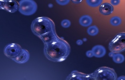
generation, the miscellaneous cells gradually reduce. Mouse embryonic fibroblasts are fusiform. However, when
connected in pieces, the cells crosslinked with each other, and the shape is not typical.
NOTES
-
The primary embryonic mouse fibroblasts isolated by the above method are not pure. The main components are
fibroblast-like cells, but there are neuro-like cells, cardiomyocytes, and some cells of unknown type.
-
After passage, the number of miscellaneous cells decreases, and the cell composition tends to be single with the
increase of passage algebra, but the proliferation ability of cells decreases, and the proliferation of cells
stops after 3-5 generations. The feeder cells used in the experiment should be passed within 3 generations.
-
In addition, the gestational age of the embryonic mice used for isolation also influences the quality and purity
of the isolated cells.
-
MEFs should be passed at a suitable density, neither too dense nor too thin. MEFs with a suitable density can be
passed at a ratio of 1 to 3.
-
In general, primary cells isolated from tissues can only be a passage through 2-3 times. This is one reason why
the method is limited.
RELATED PRODUCTS & SERVICES