Transwell Migration and Invasion Assays
Cell migration and invasion are involved in embryonic development, immune response, and many pathological processes such as cancer metastasis and inflammation. Therefore, methods to study cell migratory behavior are very useful research tools for a wide range of disciplines in biomedicine.
The most widely accepted cell migration technique is the Boyden Chamber assay. The classic transwell migration detection system uses a hollow plastic chamber sealed at one end with a porous membrane. This chamber is suspended over a larger well which may contain medium and/or chemo-attractants. Cells are placed inside the chamber and allowed to migrate through the pores to the other side of the membrane. Migratory cells are then stained and counted. The transwell migration and invasion assays can be used to analyze the ability of individual cells to directionally respond to various chemo-attractants whether they are chemokines or growth factors. They can also be used to identify and characterize the key regulatory factors that affect cell migration.
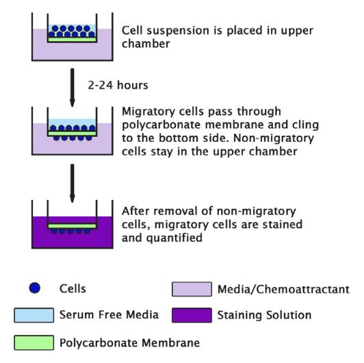 Figure 1. Schematic diagram of cell migration assay.
Materials and Equipment
Cell of interest
Trypsin-EDTA solution
Fetal bovine serum (FBS)
Phosphate buffered saline (PBS)
Serum-free cell culture media
Ethanol
Matrigel
Crystal violet
Chemo-attractant
Centrifuge
Cell culture incubator
24-well transwell inserts
Cell Preparation
- In a sterile environment, detach cells from the tissue culture plate using a non-enzymatic cell disassociation buffer or 0.25% trypsin-EDTA solution, centrifuge and aspirate the existing media leaving the pelleted cells.
- Resuspend cells in serum-free cell culture media.
Note: 1) 0.25% trypsin-EDTA may affect cell migration and invasion due to the cleavage of various receptors on the cell surface. 2) Further tests may be needed to find a seeding density that provides the best results. Typically 1 x 10⁶ cells/mL is a good starting point for most cell types when using the 24-well transwell insert. 3) There is a range of transwell insert sizes and the number of cells added should be adjusted for accordingly.
Transwell Coating (Optional)
The transwell migration assay can be easily modified to perform the cell invasion assay. To do this, add extracellular matrix (ECM) materials on top of the transwell membrane and then add cells on top of the ECM.
- Matrigel is thawed and liquefied on ice, and then 30-50 µL of matrigel is added to a 24-well transwell insert and solidified in a 37°C incubator for 15-30 minutes to form a thin gel layer.
- Cell solution is added on top of the matrigel coating to simulate invasion through the extracellular matrix.
Note: There are distinct differences between the transwell cell migration and the transwell cell invasion assays. The transwell cell migration assay measures the chemotactic capability of cells toward a chemo-attractant. The transwell cell invasion assay, however, measures both cell chemotaxis and the invasion of cells through extracellular matrix, a process that is commonly found in cancer metastasis or embryonic development.
Plating Cells
- Plate 100 μL of cell solution on top of the filter membrane in a transwell insert and incubate for 10 minutes at 37°C and 5% CO2 to allow the cells to settle down.
Note: The particular pore size of the transwell membrane used is dependent on the individual size of the cells in the sample. For example, if the cells are too small they may pass through the pores without the need to facilitate migration or if they are too large they may not fit through the pores. There are several transwell inserts with pore sizes that range from 3 µm, 5 µm, and 8 µm for cell migration assays. - Using a pipette, very carefully add 600 μL of the desired chemo-attractant into the bottom of the lower chamber in a 24-well plate.
Note: 1) Add the chemo-attractant without moving the transwell insert and avoid generating bubbles. 2) Making sure the chemo-attractant liquid in the bottom well makes contact with the membrane in the upper well to form a chemotactic gradient. - Incubate cultures for 4 to 24 hours depending on cell type and the chemo-attractant being used.
Note: For adherent cells, the migrated cells will attach to the other side of the membrane. The quantification of migrated cells can be performed in a sterile environment. For non-adherent cells, the migrated cells will drop into the media in the lower chamber. The number of migrated cells can be counted by using hemocytometer or flow cytometer.
Staining Cells
- Remove the transwell insert from the plate. Use a cotton-tipped applicator as many times as needed to carefully remove the media and remaining cells that have not migrated from the top of the membrane without damaging it.
- Add 600-1,000 μL of 70% ethanol into a well of a 24-well plate. Place the transwell insert into the 70% ethanol for 10 minutes to allow cell fixation. Remove transwell insert from the 24-well plate and use a cotton-tipped applicator to remove the remaining ethanol from the top of the membrane. Allow the transwell membrane to dry (typically 10-15 minutes).
- Add 600-1,000 μL of 0.2% crystal violet into a well of 24-well plate and position the membrane into it for staining. Incubate at room temperature for 5-10 minutes.
- Gently remove the crystal violet from the top of the membrane with a pipette tip or cotton tipped applicator. Very carefully, to avoid washing off fixed cells, dip the membrane into distilled water as many times as needed to remove the excess crystal violet. Allow the transwell membrane to dry.
- View underneath an inverted microscope and count the number of cells in different fields of view to get an average sum of cells that have migrated through the membrane toward the chemo-attractant and attached on the underside of the membrane.
Cell Services
Cell Line Testing and Assays
Explore Other Options