3D Angiogenesis Assay
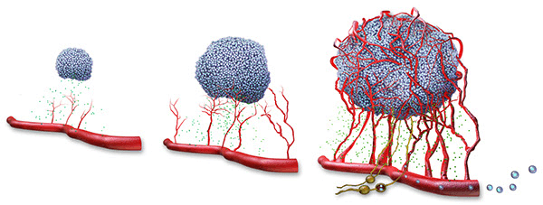
Angiogenesis, a pivotally natural process in the development of cancer malignancy, is a key step in the tumor progression towards an aggressive, metastatic state with poor response to therapies. A well-established vascular network allows tumors to overcome growth restrictions of hypoxia and nutrient deprivation and provides paths for tumor cell dissemination. During the process of angiogenesis, endothelial cells are activated, and recruited. Then these cells proliferate to organize into new 3D capillary networks and degrade their surrounding extracellular matrix to promote tumors growth and metastatic potential. The researches based on 2D monolayer cell cultures have shown limitations on their translation/extension to clinical studies because they were fail to mimic the tumor microenvironment and heterogeneity. However, multicellular tumor spheroids recapitulate the 3D architecture of tumor tissues and cell–cell interactions between tumor cells and the endothelial cells. By using 3D angiogenesis assay, the potential of tumor vascularization can be better assessed; the effect of inhibitors or inducers can also be investigated.
Creative Bioarray 3D angiogenesis assay service advantages
- Test your compounds by choosing cells from our comprehensive human and animal cell bank or sending your own cells.
- Use models that capture the interaction between supporting cells and tumors cells.
- Use multi-image read-outs for the process of angiogenesis to carry out the kinetic assays.
- Multiple cell types can be considered and introduced into the 3D systems based on the bio-relevance of each tumor models.
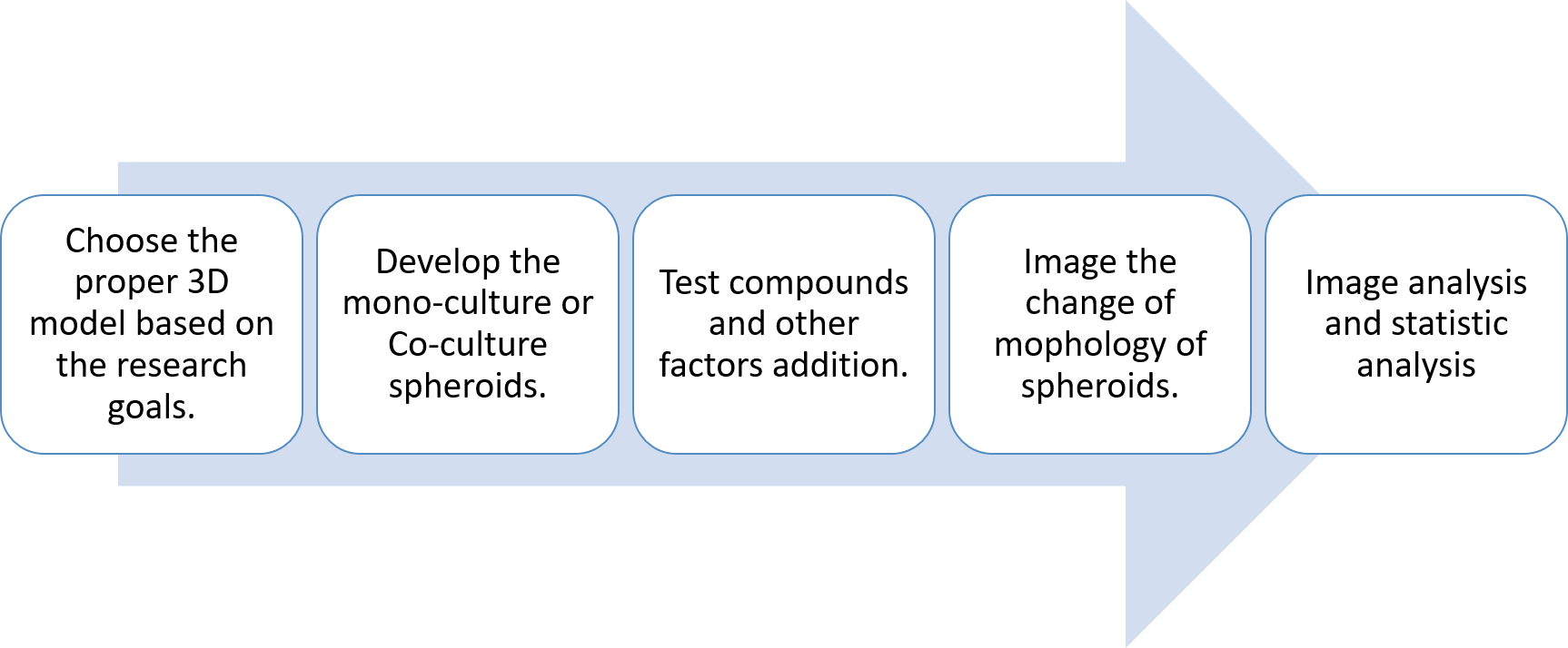
Workflow
Applications
The potential for tumor angiogenesis is often associated with the migration of endothelial cells into tumor spheroids or the formation of vascular networks within spheroids. Creative Bioarray provides different types of in vitro angiogenesis services to fulfill our customers’ demand on testing the effect of a compound for its inhibitory or inducible potential. Our characterization of angiogenesis has been established by using real-time tube formation assay or sprouting assay on 3D matrix.
Study examples
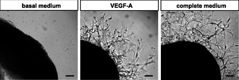
Figure 1 Representative micrographs of angiogenic outgrowth of different treatments.
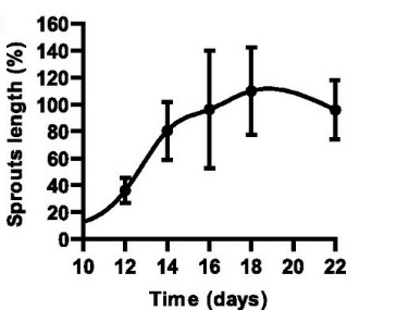
Figure 2 Time course of angiogenic outgrowth lengths in complete medium.
Quotations and ordering
Our customer service representatives are available 24hr a day!
References
- Upreti, M., et al. Tumor-endothelial cell three-dimensional spheroids: new aspects to enhance radiation and drug therapeutics. Translational oncology. 2011, 4.6: 365IN3-376.
- Blacher, S., et al. Cell invasion in the spheroid sprouting assay: a spatial organisation analysis adaptable to cell behaviour. PloS one. 2014, 9.5: e97019.
- Katt, M. E., et al. In vitro tumor models: Advantages, disadvantages, variables, and selecting the right platform. Frontiers in bioengineering and biotechnology. 2016, 4.
- Baker, M., et al. Use of the mouse aortic ring assay to study angiogenesis. Nature protocols. 2012, 7.1: 89-104.
