In Vitro Wound Healing Assay
In vitro wound healing assay is used to study the ability of cells to initiate migration in a confluent culture once a denuded area is created. This method has been used for more than 40 years and is very useful in characterizing a number of factors involved in cell migration, including the role of ECM proteins, the role of cell-cell connections, and the role of various intracellular proteins in mediating cell orientation. In this assay, a confluent monolayer of cells is "scratched" away, and cell migration is measured on the basis of the time it takes for the bordering cells to refill the denuded area. The wound healing assay is particularly relevant to the healing of the endothelium that occurs in vivo and is a relatively simple, straightforward method to study endothelial cell migration that can be performed with tools readily available in most cell biology laboratories.
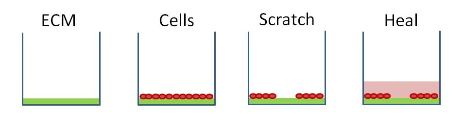 Figure 1. Basic steps involved in a wound healing assay.
Materials and Equipment
ECM protein
Endothelial cells
Fetal bovine serum (BSA)
Phosphate buffered saline (PBS)
Pipettes
Incubator
Centrifuge
Cell culture dish
Phase-contrast microscopy
Assay Procedure
1) Dissolve the ECM protein of interest in the appropriate buffer, typically PBS, but may vary depending on the protein.
2) Add the dissolved protein to the well to be coated at the desired concentration, typically ranging from 1 mg/mL to 50 mg/mL.
3) Incubate overnight at 4°C or 37°C on a level surface.
4) Aspirate off the remaining solution and wash twice with PBS.
5) Add 1% BSA solution and incubate for 1 h at 37°C.
Note: Because time, temperature, and concentration can alter the amount of protein bound to the culture dish, it is best to choose one time and temperature for all experiments.
6) Endothelial cells are plated on the dishes and grown to confluence. Because the wound healing is due to both cell migration and cell proliferation, actinomycin C can be added to the medium after the cells reach confluence at a concentration of 1 ng/mL to inhibit proliferation.
Note: The assay can be performed on native or transfected cells. If transfected cells are to be used, the cells should be transfected with the plasmid of interest and a reporter plasmid such as GFP and grown to confluence before scratching the monolayer.
7) Scratch a "wound" in the monolayer by dragging a p200 or p1000 pipette tip in a straight line across the monolayer.
8) Mark the location of the wound with a marker on the underside of the dish, which will make it easier to find the wound in the following steps.
9) Aspirate the media from the dish and replace with fresh, warmed media.
10) Use the markings created in step 8) as reference points to find an area of the wound that will be imaged throughout the experiment and acquire an image of the wound under phase-contrast microscopy.
11) Return the dish to the incubator for 6 h.
12) Locate the same area imaged in step 10) with the reference markings and take an image with phase-contrast microscopy.
13) Repeat steps 11) and 12) until the wound has completely filled in. This time will be based on the conditions and cell type, but typically takes approximately 24 h.
14) To quantify cell migration, the area of the initial wound is compared with the area of the healing wound at various time points after the scratch is imposed.

References
- Yarrow J. C, et al.; A high-thoughput cell migration assay using scratch wound healing, a comparison of image-based readout methods. BMC Biotechnol, 4: 21.
- Liang C, et al.; In vitro scratch assay: a convenient and inexpensive method for analysis of cell migration in vitro. Nature Protoc, 2007, 2: 329-333.
- Cory G. Scratch wound assay. Meth. Mol. Biol, 2011, 769: 25-30.
Cell Services
Cell Line Testing and Assays
Explore Other Options