Radioactive ISH Protocol for Detecting Gene Expression Patterns
GUIDELINE
Knowing the timing, level, cellular localization, and cell type that a gene is expressed in contributes to our understanding of the function of the gene. Each of these features can be accomplished with in situ hybridization to mRNAs within cells.
METHODS
Generation of radioactive riboprobes
- Generate a purified linear DNA template of your cDNA of interest either by an enzyme-restricted digest of a cloned fragment surrounded by RNA polymerase promoter sites from a plasmid DNA or by PCR of the insert attached to the RNA polymerase promoter binding sites.
- To a 0.5 ml tube, add 0.5-1 μg of purified linear DNA template, transcription buffer, DTT, RNasin, AGC nucleotide mix solution, and RNase-free water to bring the volume up to the desired amount. Then add S35-UTP and the appropriate RNA polymerase to make either antisense or sense riboprobes.
- Incubate the mixture for 1 hr in a 37°C water bath. Add another aliquot of RNA polymerase and incubate for another 1 hr.
- Add 3M sodium acetate solution (0.1-fold of total volume) and 100% EtOH (2.5-fold of total volume) into the tube to precipitate the synthesized RNA riboprobe.
- Incubate in dry ice or -80°C for 15 min or more than 3 hours to assist precipitation.
- Pellet RNA by centrifuging at 4°C and 15,000 rpm in a table-top centrifuge for 30 min.
- Remove the supernatant and wash the pellet with 70% EtOH, tap with a finger to mix the pellet well.
- Pellet RNA again by centrifuging at 4°C and 15000 rpm for 30 min.
- Remove the supernatant completely by pipetting and then add 40 μl hybridization solution. Mix well by pipetting in and out.
- Put 1 μl of the solution into 3 ml Safety-Solve solution in the scintillation vial, mix well, and measure the counts in the scintillation counter.
- Place the tube with the riboprobe at -20°C for use within one week.
Creative Bioarray Relevant Recommendations
- Creative Bioarray offers different types of ISH Probes and Custom Probes. Tell us the specific gene or the chromosome region of your interest, and the desired dye color, and we will make a custom probe for you.
Hybridization
- Calculate the amount of riboprobe and hybridization solution needed for all slides. Prewarm the riboprobe-hybridization mix to 65°C for 5 min to denature the riboprobe.
- Pipet 100 μl of riboprobe-hybridization solution in a line across the slide, and use a glass coverslip to spread it evenly over the tissue and coverslip. Place slides horizontally, facing upright into a metal rack and place the rack slowly upright into the 65°C oil bath for a minimum of 4 hr and a maximum of 16 hr. The oil creates an airtight seal around the coverslips.
- Remove metal racks from the oil bath and wipe the excess oil around the rack with tissue paper.
- Wash off oil from the covered slides and metal rack in glass or metal trays containing chloroform, two times. The 2nd chloroform wash can be used as the first for the next experiment.
- Transfer the rack with covered slides into a tray with 0.1% β-mercaptoethanol + 2xSSPE solution and move up and down for a few dips to loosen coverslips and remove the dissolve excess hybridization solution.
- Transfer the rack with covered slides to a fresh solution of 0.1% β-mercaptoethanol + 2xSSPE, and then remove coverslips with RNase-free forceps in the solution to prevent scratching tissue sections. Transfer the uncovered slides into the fresh rack in a tray horizontal slide rack holder, in a solution of 0.1% β-mercaptoethanol in 2xSSPE.
- Incubate the rack with slides in the fresh 0.1% β-mercaptoethanol + 2xSSPE solution at room temperature for 1 hr to remove excess unbound RNA probe. Discard this and the previous aqueous wash solutions, as well as coverslips, as radioactive waste.
- Transfer the rack with slides to a prewarmed 2xSSPE solution at 65 °C, add β-mercaptoethanol to a final of 0.1% concentration, and incubate at 65°C for 1 hr. Discard as radioactive waste.
- Transfer and incubate the rack with slides two times in prewarmed 0.1xSPPE at 65°C for 30 min each. The amount of radioactivity removed in this step is very small and no longer considered excessive radioactive waste after this step.
- Dehydrate the rack and slides in 70%, 95%, and 100% EtOH for 2 min each.
- Dry the slides in the hood for at least 30 min.
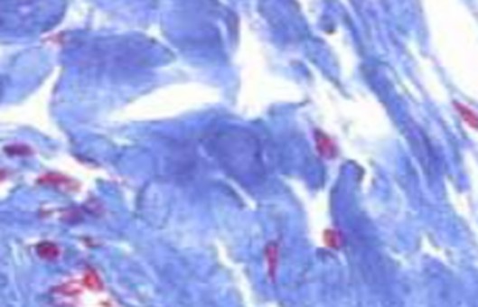
Visualization of the radioactive signal
- Place dry slides into a film cassette and in a dark room, place the x-ray film over the slides and close the cassette. Make sure that the slides are facing the emulsion side of the X-ray film. Expose the slides for 1-7 days depending on the expected abundance of the transcripts.
- Develop the X-ray film in standard developer and fixer. The hybridization signal shows up as black (exposed silver grains in the emulsion) on the film.
- Dip slides into the diluted emulsion in the 42°C water bath and dry dipped slides in a closed light tight container overnight in the dark room, or in an oven at 37°C for 2-3 hours, with lights off.
- Transfer the slides into racks slots in black boxes containing desiccators, being careful of the slides not touching each other and thus creating artifacts. Seal the edges of the boxes with black electrical tape slowly to prevent static-induced light sparks and then wrap the boxes in aluminum foil. Store the boxes at 4°C for several days to weeks (signal from 1 day on x-ray film is similar to 5 days under emulsion).
- Warm the slide boxes to room temperature for 1 hr.
- In the darkroom, remove the rack with slides (or place slides in a metal rack if using boxes with slide slots) from the boxes and develop them in the Kodak D-19 developer at 16°C for 3.5 min.
- Wash the developed slides in tap water at room temperature for 1 min.
- Incubate the slides twice in a fixer at 19°C for 6 min each. Lights can be turned on during the second fixer incubation.
- Wash the slides in running water at room temperature for at least 30 min and scrape the emulsion from the back side of the slide while 'wet' with a razor blade to prevent scratching the glass.
- Stain tissue with 0.3% cresyl violet in tap water for 5 min.
- Wash extra cresyl violet solution in fresh tap water for 15 dips.
- Dehydrate the slides for 15 dips in each alcohol solution, 50%, 70%, 95%, 95%, 100%, and 100% EtOH.
- Incubate the slides in xylene for 5 min at room temperature twice.
- Coverslip with Permount medium on the slide and dry the covered slide in the hood overnight (>16 hr); it will take several days before the glue is sturdy enough to clean the slides further.
NOTES
- The reason for using a metal rack in most of the steps is the use of chloroform washes and xylene. Both organics melt many kinds of plastics. Glass and some kinds of plastics are resistant to these organics.
- Folded tissue could be misleading for gene expression results on X-ray film, leading to a region with a darker signal. To determine if the tissue is folded, examine non-stained sections under darkfield or cresyl violet stained sections under brightfield.
RELATED PRODUCTS & SERVICES

Reference
- Chen CC, Wada K, Jarvis ED. (2012). "Radioactive in situ hybridization for detecting diverse gene expression patterns in tissue." J Vis Exp. (62), 3764.