Radioactive ISH Protocol for Animal Chromosomes
GUIDELINE
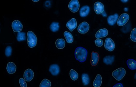
- Although largely replaced by the use of fluorescent in situ hybridization (FISH) in animal and human molecular cytogenetics, the technique of radioactive in situ hybridization (RISH) still has some uses. Using practicable exposure times for autoradiographs of 3-4 weeks, RISH is approximately 50 times more sensitive than FISH using biotin- or digoxygenin-labeled probes.
- In addition, the sensitivity of RISH can be increased with longer exposures, in a roughly linear fashion until the silver bromide grains in the emulsion approach saturation over the target.
METHODS
Acetylation of slides
- Place the slides in 500 mL of 0.1 M triethanolamine-HCl. Add 2.5 mL of acetic anhydride drop by drop, stirring continuously. Leave still for 10 min.
- Rinse slides in four changes of 2×SSC, dehydrate through an ethanol series, air dry, and perhaps store the slides.
- Make up to 20% w/v dextran sulfate in deionized formamide and heat to 70°C. Mix frequently over the next few hours, using a vortex mixer.
Preparation of hybridization mix
- Centrifuge the alcohol-precipitated mixed DNA(s) for 15 min at 17,000g (maximum speed of a microfuge).
- Pour off the supernatant and check that a pellet has been left behind.
- Dry the mixed DNA at room temperature or briefly at 37°C. If the tube contains the exact amount of mixed DNA for hybridization, then leave it dry.
- If required by the experimental design, resuspend the mixed DNA in H2O at a concentration of 5 ng/mL of the probe. Preferably leave overnight on a shaker, or heat to 70°C for a few minutes and vortex mix.
- The hybridization mix should contain the following, added in the order A-C. A. 3 vol, mixed DNA to yield a final concentration of the probe DNA in the hybridization mix as 200 ng/mL. B. 2 vol, 10×SSCP. C. 5 vol, 20% dextran sulfate in deionized formamide at 70°C.
- The actual volumes used can be calculated from the amount required to be placed on each slide under coverslips of different sizes.
- If a measured amount of the probe DNA has been left dry, then simply add the necessary volume of water, heat briefly to 70°C, and mix well.
Hybridization
- Denaturation of chromosomal DNA and the probe DNA.
- Clean the required number of coverslips by dipping them in 70% ethanol and wiping them dry.
- Place the required volume of hybridization mix to suit the size of the coverslip on each slide and add the coverslip, leaving a 3-4 mm gap at the non-frosted end (where high-power objective lenses cannot safely reach and the emulsion will be too thick).
- Seal the coverslip with generous quantities of rubber cement, containing a hydrocarbon solvent that is not miscible with water. Allow the cement to dry, for swiftness at 37°C.
- Incubate the slides flat in a slightly humid environment (e.g., plastic slide box with lightly dampened paper on the bottom) overnight for a maximum of 16 h, at 37°C for short unique probes, 42°C for long (+1500 bp) or repetitive probes.
Post hybridization
- Make up 200 mL, or more, of 50% deionized formamide/2×SSC and add 40-50 mL to at least four Coplin jars.
- Prewarm, to 2°C above the hybridization temperature, all but one of the three Coplin jars containing 50% deionized formamide/2×SSC, plus five jars containing 2×SSC.
- Remove the rubber cement from the slides using fine forceps and lift off the coverslips.
- Rinse briefly in the Coplin jar containing 50% deionized formamide/2×SSC at room temperature.
- Rinse the slides for 3 min, with agitation, in each of the eight Coplin jars.
- Dehydrate through 35%, 70%, 95%, and 100% ethanol, 2 min each, and dry.
Staining, viewing, and photographs
- Stain the slides in a Coplin jar for 20 min. Rinse twice, very briefly, in the pH 6.8 buffer.
- For permanent mounting, soak off the immersion oil with xylenes and mount a coverslip with Depex.
- Focus mainly on the level of the signal grain(s). Take several shots, going from the chromosomes in focus, through average focus, to grains only in focus.
Creative Bioarray Relevant Recommendations
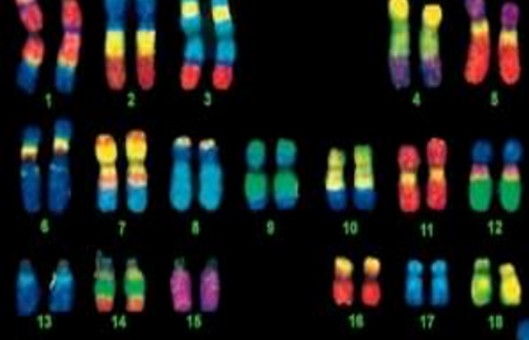
- Chromosomal abnormalities and variations are contributing factors to infertility, reduced fertility, malformations, intersex and fetal death. Creative Bioarray provide chromosome analysis for animals, gametes, embryos and stem cells.
NOTES
- At all stages practice normal procedures of sterility and no contamination for molecular biology.
- For different coverslips, the appropriate amount of probe is to be added.
RELATED PRODUCTS & SERVICES
Reference
- Darby, Ian A. (1999). "In Situ Hybridization Protocols." (163), 29-50.