Protocol for Staining FFPE Tissue with Streptavidin-HRP/Biotin
GUIDELINE
Staining FFPE (formalin-fixed paraffin-embedded) tissue with streptavidin-HRP (horseradish peroxidase) and biotin involves utilizing the strong affinity of biotin for streptavidin and the enzymatic activity of HRP for visualizing specific targets within the tissue. Here we provide a streptavidin-HRP/biotin staining protocol for FFPE tissue.
METHODS
- Place the slide in Deionized Water (DI). Perform antigen retrieval. If the slide does not require antigen retrieval, then place the slide in DI or wash buffer such as PBST: Phosphate Buffered Saline (1×PBS, pH 7.2 with 0.05% Tween 20) or TBST: Tris Buffered Saline (1X TBS, pH 7.6 with 0.05% Tween 20). Wash the slide twice for three minutes each.
- Tap off the excess buffer and place the slides on the immunostaining tray. Gently dry around the tissue section before each incubation. Do not allow the tissue to dry out during the entire immunostaining procedure.
- Incubate the tissue section with a protein block for 10 minutes. Apply enough reagent to cover the sample. Tap off the protein block; do not wash the slide.
- Use an appropriate protein block to reduce non-specific binding that may result from hydrophobic or ionic interactions.
- Incubate the section with an avidin-biotin block. First, incubate with the avidin solution for 10 minutes. Wash the slide twice for three minutes each. Then, incubate with the biotin solution for 10 minutes. Wash the slide twice for three minutes each.
- Incubate the tissue section with the primary antibody and control for 30 minutes to 2 hours at room temperature or overnight at 4˚C. Adjust the incubation time as needed. Wash the slide twice for three minutes each.
- Incubate the tissue section with any necessary reagents to block endogenous molecules, such as endogenous peroxidase or phosphatase, with the appropriate blocking reagent(s) for 10 minutes. Wash the slide twice for three minutes each.
- Each staining system utilizes an enzyme to catalyze the chromogen color development solution. However, the enzyme employed may also be naturally occurring in the tissue specimen. For example, peroxidase is present in red blood cells, muscles, kidneys, etc. If utilizing a horse-radish peroxidase enzyme, then block endogenous peroxidase with a hydrogen peroxide solution to prevent non-specific color development.
- Incubate the tissue section with the corresponding biotinylated secondary antibody for 30 minutes. Wash the slides twice for three minutes each.
- Incubate the tissue section with the streptavidin-peroxidase conjugate for 30 minutes. Wash the slides twice for three minutes each.
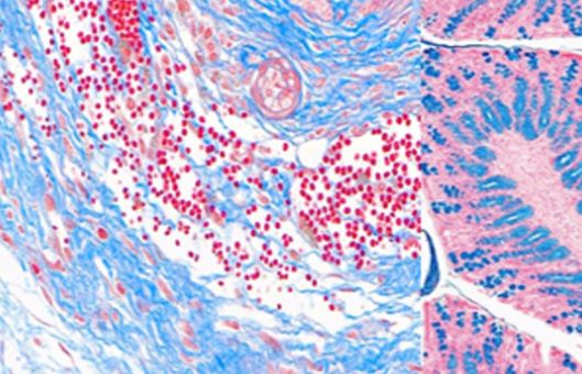
- Select the correct chromogen, such as DAB (3, 3' Diaminobenzidine) with the peroxidase enzyme. Incubate the tissue section with the chromogen for 5-10 minutes. Refer to the chromogen data sheet instructions. Wash the slide with DI for 3-5 minutes.
- Counterstain the tissue section with hematoxylin for 2-5 minutes. Wash the slide with DI for 3-5 minutes. Rinse with tap water or an alkaline solution to enhance the counterstain.
- The cell nuclei are generally a purple/blue color after the hematoxylin incubation (low pH) and upon incubation with a basic solution the nuclei turn blue.
- Dehydrate and clear the tissue section.
- Coverslip the slide with an appropriate mounting media.
Creative Bioarray Relevant Recommendations
- Creative Bioarray offers a variety of fluorescent dyes and special dyes to help you better understand molecular biology.
- We also offer the most comprehensive list of high-quality and affordable FFPE cell pellets to best fit your specific research objectives, including but not limited to the following.
| Animal Cells | Brain Tumors | Breast Tumors |
| Cervical Tumors | Colon Tumors | Kidney Tumors |
| Leukemia | Liver Tumors | Lung Tumors |
| Lymphoma | Melanoma | Ovarian Tumors |
| Pancreatic Tumors | Prostate Tumors | Stomach Tumors |
NOTES
- Properly block endogenous peroxidase activity and nonspecific binding sites to minimize background staining. Use appropriate blocking reagents and buffers to prevent nonspecific interactions with the tissue and antibodies.
- Ensure the selection of high-quality Streptavidin-HRP conjugates and biotinylated secondary antibodies that are specific to the target antigens and compatible with the tissue species.