MTT Analysis Protocol
GUIDELINE
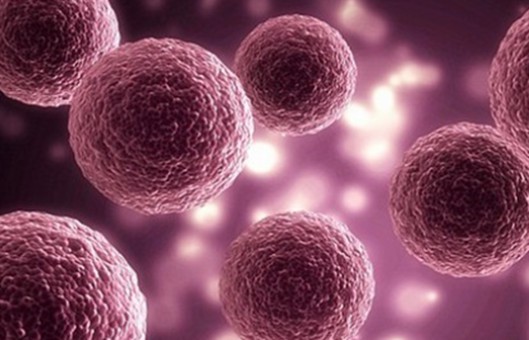
This experiment describes a method to detect cell survival and growth-MTT analysis assay. The principle of the assay is that the enzyme succinate dehydrogenase in the mitochondria of living cells reduces exogenous MTT to water-insoluble blue-violet crystals of Formazan and deposits them in the cells, whereas dead cells do not have this function. Dimethyl sulfoxide (DMSO) dissolves Formazan in the cells, and its light absorption value is measured at 490 nm with an enzyme marker, and the amount of MTT crystals formed is proportional to the number of cells within a certain cell number range. According to the measured absorbance value (OD value), to determine the number of living cells, the larger the OD value, the stronger the cell activity (if it is to measure the drug toxicity, it means that the drug toxicity is smaller).
METHODS
- Trypsin digestion of log phase cells is terminated and collected by centrifugation to make cell suspension. The concentration is adjusted to 5-10×104/ml for cell counting.
- The cell suspension is prepared and gently mixed. 100 μl per well is added so that the density of cells to be tested is 5000-10000/well (edge wells are filled with sterile PBS).
- Put the inoculated cell culture plate into the incubator and incubate it until the cell monolayer spreads over the bottom of the wells (96-well flat-bottomed plate), and add the drug with the concentration gradient. Generally, 5-7 gradients, 100 μl per well, set up 3-5 compound wells. It is recommended to set up 6, otherwise, it is difficult to reflect the real situation.
- 5% CO2, incubate at 37°C for 16-48 hours and observe the effect of the drug under the inverted microscope.
- Add 10 μl MTT solution (5 mg/ml, i.e. 0.5% MTT) to each well, and continue incubation for 4 h. If the drug and MTT can react, centrifuge and discard the culture solution first, and then add the MTT-containing culture solution after carefully rinsing it with PBS 2-3 times.
- Terminate the incubation and prepare to dissolve the crystals. MTT is added to incubate for 4 h, the crystals can be fully formed. 150 μl of dimethyl sulfoxide is added to each well and placed on a shaker with low-speed shaking for 10 min to fully dissolve the crystals. The absorbance value of each well is measured at OD490 nm of the enzyme immunoassay detector.
- Zeroing wells (culture medium, MTT, dimethyl sulfoxide) and control wells (cells, same concentration of drug dissolution medium, culture medium, MTT, dimethyl sulfoxide) are also set up.
Creative Bioarray Relevant Recommendations
- Creative Bioarray provides SuperQuick® cell proliferation kit I (MTT) which is a colorimetric assay for the non-radioactive quantification of cellular proliferation, viability, and cytotoxicity. The sample material is either adherent or suspension cells cultured in 96-well microplates.
- We also offer a wide selection of cell viability assays for drug screening and cytotoxicity tests of chemicals, including MTT/MST/WST-8/WST-1/CKK-8 assays, trypan blue assays, calcein-AM assays, fluorescent reactive dye assays, single/two color viability assays, and microplate cell viability assays.
NOTES
- Because the cells continue to settle even after mixing, the inoculation process should be repeated several times. For example, mixing every 6 wells is added to ensure that the density of inoculated cells is the same from one well to another, which is crucial for the results of MTT. In addition, the number of cells should also be determined according to the characteristics of the cells themselves, such as the growth rate.
- In principle, the drug can be added after the cells are attached to the wall, for two hours, or half a day. But we often lay the plate in the afternoon of the previous day and add the drug in the morning of the next day.