ISH Protocol to Polytene Chromosomes
GUIDELINE
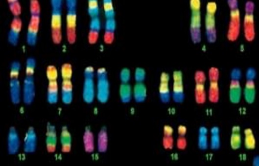
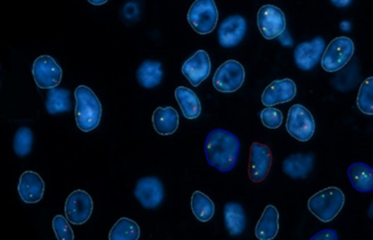
- The power of in situ hybridization is due largely to the scale of polytene chromosomes and consequently the high degree of resolution they offer the researcher. The use of radiolabeled probes has now been largely superseded by nonradioactive signal detection systems, generally using biotin- or digoxygenin-substituted probes, which offer still greater resolution because there is less scatter of signal with immunochemical and immunofluorescent detection than with silver grains.
- The utilization of in situ hybridization technology is of particular interest to those engaged in chromosome walking or genome mapping projects, in which it is essential to check all clones along a chromosome walk by in situ hybridization to identify clones containing repetitive DNA and to avoid the isolation of clones derived from regions outside that of interest.
METHODS
Pretreatment and denaturation of chromosomes
- Place the slides directly in gently boiling 10 mM Tris-HCl, pH 7.5, for 2 min.
- Quickly transfer the slides to cold 70% ethanol, and dehydrate them through alcohol.
Preparation of labeled probe
- Place 5 μL oligo labeling buffer and 1 μL of 1 mM fluorescein-12-dUTP in a microcentrifuge tube.
- Boil 100-500 ng of DNA in 20 μL of water or TE for 3 min, then add 18 μL to the tube.
- Add 1 μL (5 U) of Klenow fragment of DNA polymerase I and incubate the reaction at room temperature for 1 h overnight.
- Ethanol precipitate the labeled DNA and resuspend it in 50 μL of sterile distilled water to which 50 μL of 2×hybridization solution is added.
Hybridization
- Boil the probe for 3 min by suspending the tube in boiling water, and quench it on ice. Check the volume after boiling, and restore to the initial volume with sterile distilled water.
- Pipette 20 μL of the probe onto the chromosomes, then cover the chromosomes and probe with a clean siliconized coverslip. Avoid trapping air bubbles underneath the coverslip. There is no need to seal the coverslip.
- Place the slides in a humid box (a plastic box lined with moist tissue) to prevent evaporation from the preparation, seal the lid, and place in a 58°C incubator overnight.
- Remove the slides from the humid box. Dip them in 2×SSC to allow the coverslip to slide off, then wash them in 2×SSC, 53°C, for 1 h. Three changes to the wash solution should be made.
Signal Detection
- The slides should be taken from the final wash and passed through two 5 min washes in PBS.
- Wash the slides for 2 min in PBS-TX.
- Rinse in PBS. Do not allow the slides to dry out during signal detection.
- Drain the slide and mount it in a mounting medium, under a siliconized coverslip. The mounting medium contains propidium iodide to stain the chromosomes.
- Seal the coverslip with nail varnish. These preparations do not last a long time but will keep for several weeks at 4°C in the dark if sealed as described. They are best examined using a microscope with a facility for image merging. A confocal microscope is ideal.
Creative Bioarray Relevant Recommendations
- Creative Bioarray offers a wide range of ISH products and services, from ISH Probes and ISH Reagents to In Situ Hybridization (ISH) & RNAscope Service and Digital ISH Image Quantification and Statistical Analysis. In addition, we provide chromosome analysis for animals, gametes, embryos and stem cells. We can customize the standard cytogenetic analysis to meet your needs.
NOTES
There are many ways in which polytene chromosomes can be prepared, differing mostly in the manner by which the chromosomes are spread and squashed. Allowing the coverslip to slip sideways when spreading causes the chromosome arms to stretch. Overstretched chromosomes can make an analysis of the in situ hybridization difficult. Make sure the slides and coverslips are clean, especially of lint from the tissue paper used to clean them, thus, only lint-free tissue paper should be used.
RELATED PRODUCTS & SERVICES
Reference
- Asp J, et al. (2006). "Nonradioactive in situ hybridization on frozen sections and whole mounts." Methods Mol Biol. 326, 89-102.


