Identification of Lymphatic Glycocalyx by TEM and IHC
Scientific Reports. 2023 Feb 21; 13 (1): 3022.
Authors: Gianesini S, Rimondi E, Raffetto JD, Melloni E, Pellati A, Menegatti E, Avruscio GP, Bassetto F, Costa AL, Rockson S.
INTRODUCTION
- Blood flow is translated into biochemical inflammatory or anti-inflammatory signals based on shear stress type, utilizing sensitive endothelial receptors. Recognition of the phenomenon is of paramount importance for enhanced insights into the pathophysiological processes of vascular remodeling.
- The endothelial glycocalyx is a pericellular matrix, identified in both arteries and veins, acting collectively as a sensor responsive to blood flow changes. Venous and lymphatic physiology are interconnected. However, to our knowledge, a lymphatic glycocalyx structure has never been identified in humans.
METHODS
- Transmission electron microscopy (TEM). The specimens were divided into two fragments, one for histological analyses by light microscopy and the other for morphological analyses by transmission electron microscopy (TEM). For TEM analyses, the lymphatic vessels and veins were fixed in 2.5% glutaraldehyde in 0.1 M cacodylate buffer (pH 7.4), containing 0.05% (w/v) Alcian Blue 8GX, at 4°C over-night and post-fixed in 2% buffered osmium tetroxide for 1 h. The specimens were then dehydrated with graded concentrations of acetone and embedded in epoxy resin.
- Immunohistochemistry (IHC). For histological analyses, the lymphatic and venous samples were fixed in formalin 10% for 24 h at 4°C, dehydrated through an alcohol series, and then paraffin-embedded. Five-μm-thick sections were cut from paraffin blocks. The sections were stained with the primary anti-podoplanin, anti-glypican-1, anti-agrin, anti-brevican antibodies, and the anti-mucin-2 antibody. And then counterstained with the anti-rabbit HRP-DAB tissue staining kit. A negative control was obtained in each slide by carrying out the immunohistochemistry staining procedure without using the primary antibody.
- Browse our recommendations
We provide a range of high-quality products and services for our customers' research, including but not limited to the products in the table below.
| Service Types | Description | Recommended Products |
| Transmission Electron Microscopy Service | It is a technique in which high-energy electrons are transmitted through electron-transparent samples. | Transmission Electron Microscopy (TEM) Service, Particle Size Analysis, SuperCult® Alcian Blue Staining Kit… |
| Immunohistochemistry Service | IHC, by exploiting the principle of antibodies binding specifically to antigens in biological tissues, is a routine method to detect antigens in cells of a tissue section. | Immunohistochemistry (IHC) Service, Steady DAB/Plus… |
RESULTS
- After fixation, the wall thickness of the vein and the lymphatic samples measured X10 along the vessel transverse section were 397.3 ± 208.3 mm, and 51.3 ± 12.6 mm, respectively. TEM analysis confirmed the presence of a GCX-like structure along both the vein and lymphatic endothelium. The mean height of this structure measured 17X along the vein section and 25X along the lymphatic transverse section was 66.94 ± 18.38 nm in the vein, and 49.26 ± 11.30 nm in the lymphatic sample (p < 0.001).
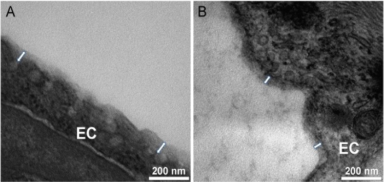 Fig. 1 Glycocalyx-like structure assessment of lymphatic vessels.
- Immunohistochemical analyses were performed in various sections of the lymphatic vessel and vein utilizing antibodies to anti-podoplanin, anti-glypican 1, and anti-mucin-2 (scale bar, 50 μm). Both venous and lymphatic immunohistochemistry identified glypican-1 and mucin-2. All the results were confirmed by the negative control performed by the secondary antibody. Podoplanin, widely recognized as a lymphatic tissue-specific marker, was identified only in the lymphatic vessel.
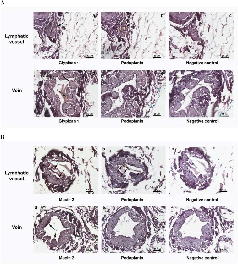 Fig. 2 Immunohistochemistry showing lymphatic glycocalyx-like structure components.
SUMMARY
- The present investigation provides the first in human identification and characterization of a GCX structure also present in the lymphatic system, providing a comparative analysis of ex vivo samples of lymphatic and venous tissue obtained from the same subject.
- The study provides fundamental information and technical applications for further assessments in the different parts of the lymphatic systems that may be both physiological and pathological conditions and potentially provide translational resources that will inform the future management of the extremely relevant clinical issue of lymphedema.
RELATED PRODUCTS & SERVICES
Reference
- Gianesini S, et al. (2023). "Human collecting lymphatic glycocalyx identification by electron microscopy and immunohistochemistry." Sci Rep. 13 (1), 3022.