GUIDELINE
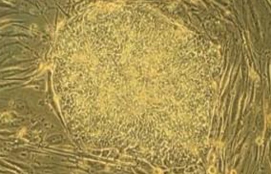
- The Colony-Forming Cell (CFC) assay, also known as the Colony-Forming Unit (CFU) assay, is the current gold standard for in vitro testing of hematopoietic stem/progenitor cell function. Hematopoietic progenitor cells proliferate and differentiate during culture to produce mature blood cell colonies, and the function and quality of hematopoietic cells are identified and analyzed based on the number and type of cell colonies (the latter judged by morphology and cell phenotype). CFU colonies are generally divided into erythroid colony-forming units (CFU-E), blast erythroid colony-forming units (BFU-E), granulocyte colony-forming units (CFU-G), macrophage colony-forming units (CFU.M), granulocyte/macrophage (CFU-GM) and mixed cell lineage colony-forming unit (CFU-GEMM).
- In the CFU colony assay, two points need to be considered in focus. One is the medium used to maximize the growth and division of hematopoietic cells and their progeny within the colonies. Two is the matrix within the medium, which must increase the viscosity of the medium, but not yet turn the medium into a solid. Therefore, the use of a semi-solid medium is critical, as the semi-solid medium allows for the formation of colonies by limiting the progeny of individual progenitor cells to a fixed location, which allows for the identification and counting of different colony morphologies.
METHODS
- Collect blood samples in sterile tubes containing heparin sodium salt to avoid clotting.
- Remove erythrocytes by immunogenicity gradient centrifugation with compounding more (erythrocyte lysate), Hetasep, and SepMate.
- Wash cells with IMDM containing 2% FBS and adjust cell concentration.
- Count nucleated cells with methylene blue containing 3% acetic acid or count live cells using the tabletop blue exclusion method to determine cell concentration.
- Add the cells to the dispensed MethoCult (methylcellulose) semi-solid medium by vortexing and shaking.
- Insufficient cell inoculation may result in too few colonies, resulting in statistically insignificant counts or inaccurate statistics. A higher inoculation density will result in excessive nutrient depletion in the culture, accumulation of cellular metabolites, inhibition of progenitor cell proliferation, and overlapping cell colonies due to too many colonies, making it impossible to count colonies accurately.
- For a 1.1 mL culture system with two-well replicates, 0.3 mL of the adjusted concentration of cell suspension is added to a tube prefilled with 3.0 mL of MethoCul. For a 1.1 mL culture system with three well replicates, 0.4 mL of diluted cells are added to a tube prefilled with 4.0 mL of MethoCult. The medium ratio has the appropriate viscosity to ensure optimal CFU growth and morphology.
- Place the inoculated 35 mm culture and an uncovered 35 mm dish (containing 3 mL sterile water) in a 245 mm square dish. If using a SmartDish, add 4 mL sterile water to the slot between the SmartDish wells and place 4 uncovered 35 mm dishes (containing 3 mL sterile water) together in a 245 mm square dish. The use of square Petri dishes with lids helps to maintain humidity during the incubation process, prevents the medium from drying out, and also reduces contamination. Additions (e.g., copper sulfate crystals) can also be added to the water-containing dishes to prevent microbial growth.
- CFU colonies can be detected after culture for about 14 days at 37°C, with 5% CO2 and 95% humidity, and only the number of Petri dishes that can be counted within 1 hour is taken out each time.
- If the cells are cultured in a SmartDish 6-well plate, cell counting can be performed with a STEMgrid-6 counting grid. The counting grid is specifically designed to count colonies in SmartDish 6-well plates and can also be used with standard 35 mm Petri dishes.
Creative Bioarray Relevant Recommendations
NOTES
- If EDTA (ethylenediaminetetraacetic acid) and ACD (glucose citrate) are used for collection, an additional anticoagulant needs to be added to the medium during dilution and washing.
- In the case of frozen blood samples, erythrocytes have been lysed after resuscitation and cannot be removed using Hetasep.
- For serum-free culture conditions, wash with IMDM containing 25 mm HEPES.
- Cells should be counted within 10 minutes of the addition of methylene blue, or in the case of benchtop blue counting, cells should not be incubated in benchtop blue for more than 15 minutes, otherwise cell death may occur, resulting in inaccurate counts.
- After vortexing for 5 minutes, let stand at 4°C for 15-30 minutes to allow air bubbles to rise to the collar and immediately inoculate the mixed cells into Petri dishes in 6-well culture plates by inoculating the appropriate number of cells in 35 mm culture or SmartDish 6 plates.
RELATED PRODUCTS & SERVICES