- You are here: Home
- FISH Slide Making Protocol for Metaphase Spreads
Services
-
Cell Services
- Cell Line Authentication
- Cell Surface Marker Validation Service
-
Cell Line Testing and Assays
- Toxicology Assay
- Drug-Resistant Cell Models
- Cell Viability Assays
- Cell Proliferation Assays
- Cell Migration Assays
- Soft Agar Colony Formation Assay Service
- SRB Assay
- Cell Apoptosis Assays
- Cell Cycle Assays
- Cell Angiogenesis Assays
- DNA/RNA Extraction
- Custom Cell & Tissue Lysate Service
- Cellular Phosphorylation Assays
- Stability Testing
- Sterility Testing
- Endotoxin Detection and Removal
- Phagocytosis Assays
- Cell-Based Screening and Profiling Services
- 3D-Based Services
- Custom Cell Services
- Cell-based LNP Evaluation
-
Stem Cell Research
- iPSC Generation
- iPSC Characterization
-
iPSC Differentiation
- Neural Stem Cells Differentiation Service from iPSC
- Astrocyte Differentiation Service from iPSC
- Retinal Pigment Epithelium (RPE) Differentiation Service from iPSC
- Cardiomyocyte Differentiation Service from iPSC
- T Cell, NK Cell Differentiation Service from iPSC
- Hepatocyte Differentiation Service from iPSC
- Beta Cell Differentiation Service from iPSC
- Brain Organoid Differentiation Service from iPSC
- Cardiac Organoid Differentiation Service from iPSC
- Kidney Organoid Differentiation Service from iPSC
- GABAnergic Neuron Differentiation Service from iPSC
- Undifferentiated iPSC Detection
- iPSC Gene Editing
- iPSC Expanding Service
- MSC Services
- Stem Cell Assay Development and Screening
- Cell Immortalization
-
ISH/FISH Services
- In Situ Hybridization (ISH) & RNAscope Service
- Fluorescent In Situ Hybridization
- FISH Probe Design, Synthesis and Testing Service
-
FISH Applications
- Multicolor FISH (M-FISH) Analysis
- Chromosome Analysis of ES and iPS Cells
- RNA FISH in Plant Service
- Mouse Model and PDX Analysis (FISH)
- Cell Transplantation Analysis (FISH)
- In Situ Detection of CAR-T Cells & Oncolytic Viruses
- CAR-T/CAR-NK Target Assessment Service (ISH)
- ImmunoFISH Analysis (FISH+IHC)
- Splice Variant Analysis (FISH)
- Telomere Length Analysis (Q-FISH)
- Telomere Length Analysis (qPCR assay)
- FISH Analysis of Microorganisms
- Neoplasms FISH Analysis
- CARD-FISH for Environmental Microorganisms (FISH)
- FISH Quality Control Services
- QuantiGene Plex Assay
- Circulating Tumor Cell (CTC) FISH
- mtRNA Analysis (FISH)
- In Situ Detection of Chemokines/Cytokines
- In Situ Detection of Virus
- Transgene Mapping (FISH)
- Transgene Mapping (Locus Amplification & Sequencing)
- Stable Cell Line Genetic Stability Testing
- Genetic Stability Testing (Locus Amplification & Sequencing + ddPCR)
- Clonality Analysis Service (FISH)
- Karyotyping (G-banded) Service
- Animal Chromosome Analysis (G-banded) Service
- I-FISH Service
- AAV Biodistribution Analysis (RNA ISH)
- Molecular Karyotyping (aCGH)
- Droplet Digital PCR (ddPCR) Service
- Digital ISH Image Quantification and Statistical Analysis
- SCE (Sister Chromatid Exchange) Analysis
- Biosample Services
- Histology Services
- Exosome Research Services
- In Vitro DMPK Services
-
In Vivo DMPK Services
- Pharmacokinetic and Toxicokinetic
- PK/PD Biomarker Analysis
- Bioavailability and Bioequivalence
- Bioanalytical Package
- Metabolite Profiling and Identification
- In Vivo Toxicity Study
- Mass Balance, Excretion and Expired Air Collection
- Administration Routes and Biofluid Sampling
- Quantitative Tissue Distribution
- Target Tissue Exposure
- In Vivo Blood-Brain-Barrier Assay
- Drug Toxicity Services
FISH Slide Making Protocol for Metaphase Spreads
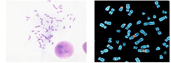
Metaphase preparation’s quality is a critical parameter for fluorescent in situ hybridization (FISH) methodologies. This protocol can be used for metaphase preparation from some kinds of cell lines. And this protocol is just for reference only. At Creative Bioarray, we can provide services for entire FISH process with unprcedented analytical accuracy, you can find more at Creative Bioarray’s Histology Services.
1) | Cells culture should be sub-confluent and in an exponentially growing phase. Change medium 4 hours prior to start of experiment. |
2) | Incubate cells with 100 μl colcemid in 5 ml of medium for 3h in order to arrest cells in the metaphase stage. |
3) | Wash cells once with PBS and then trypsinise cells as usual, collect in a 15 ml tube and spin down for 5 minutes at 800 rpm. |
4) | Remove supernatant carefully. |
5) | Snap the tube to loosen the pellet. Add 1 ml of pre-warmed (37°C) 0.56% KCl dropwise. Snap the pellet gently to get a uniform suspension. Add an additional 4 ml 0.56% pre-warmed KCl and incubate for 10 minutes at 37°C. |
6) | Centrifuge the tube at 800 rpm for 5 minutes and remove the supernatant carefully. |
7) | Snap the tube to loosen the pellet. Add 1-1.5 ml of ice-cold fixative (MeOH: Acetic acid= 3:1, prepared fresh and ice cold) to the above tube. Transfer the contents of the tube to a 1.5 ml tube. Centrifuge at 6000 rpm for 1 minute and carefully remove the supernatant. Repeat this step two to three times. |
8) | Resuspendin fixative (about 1 ml) and this is now ready to be dropped on slides. |
9) | Dip the slides in ice cold 40% methanol and allow to drip on a filter paper. With a pipette, drop 25-35 μl of cell suspension on the slide from a height about 10 cm. As the fixative gradually evaporates, the surface of the slide becomes grainy. At this moment, place the slide face down into the steam of hot water bath (75°C) for 5-10s and then dry quickly by placing the slide on a metal plate. |
10) | Incubate at 65°C overnight for aging. The slides can be stored dipped in 100% EtOH at -20°C until use. |
For research use only. Not for any other purpose.