iPSC Generation Using microRNA miR-302-based Reprogramming Method
MiR-302, the most abundant microRNA (miRNA) in human ESCs (hESCs), not only silences the expression of lysine-specific histone demethylase 1/2 (LSD1/2, AOF2/1 or KDM1/1B) and DNA (cytosine-5-)-methyltransferase 1 (DNMT1) to induce global DNA demethylation but also stimulates cellular Oct4-Sox2-Nanog co-expression to promote the full reprogramming of somatic cells into ESC-like iPSCs. Hence, miR-302 can replace all four previously defined factors (either Oct4-Sox2-Klf4-c-Myc or Oct4-Sox2-Nanog-Lin28) for iPSC induction.
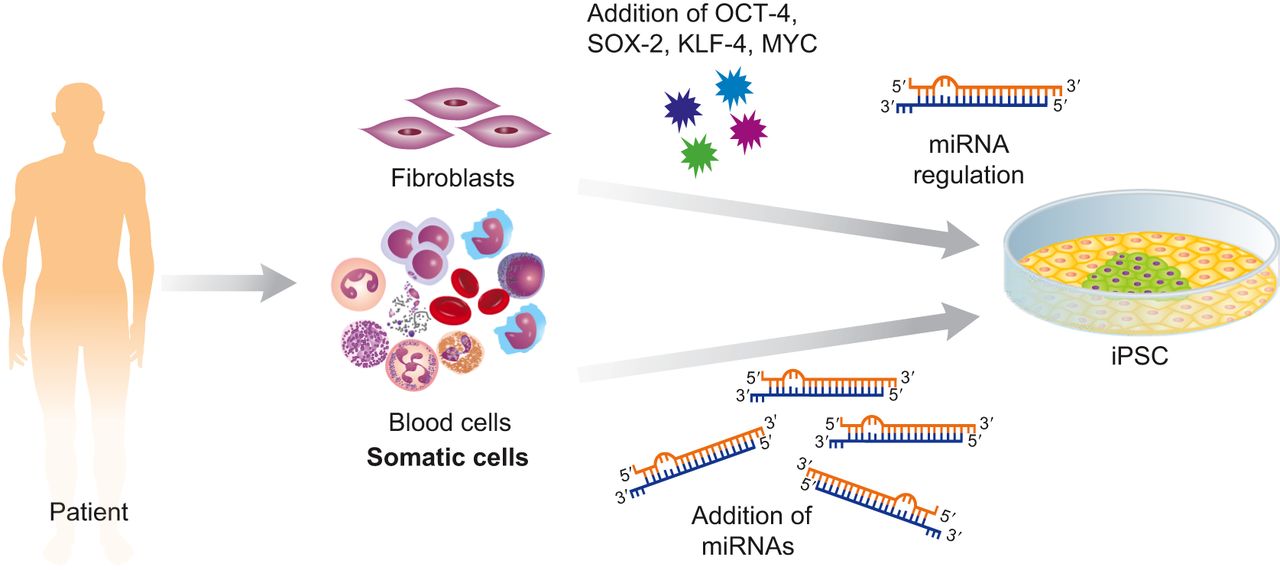 Figure 1. Somatic cells can be reprogramming by using the transcription factors OCT-4, SOX-2, KLF-4, and MYC or by the addition of miRNAs alone.
Materials and Equipment
| 70-bp cut-off purification filter | 100-bp cut-off purification filter |
| Sterilized platinum loop or pipet tip | Incubation chambers |
| Electroporator | Microcentrifuge |
| G418 | Autoclaved ddH2O |
| T4 ligase | Polysomal or liposomal reagents |
| 10X digestion buffer | Electroporation buffer |
| 2X hybridization buffer (200 mM KOAc, 60 mM HEPES KOH, 4 mM MgOAc (pH 7.4 at 25°C)) | 10X ligation buffer (660 mM Tris-HCl (pH 7.5 at 20°C), 50 mM MgCl2, 50 mM dithioerythritol, and 10 mM ATP) |
| Luria Bertani (LB) agar plate (50 mg/mL kanamycin for pSpRNAi-RGFP or 100 mg/mL ampicillin for pLVXAcGFP-N1 clone selection) | Low salt LB broth (50 mg/mL kanamycin for pSpRNAi-RGFP or 100 mg/mL ampicillin for pLVX-AcGFPN1 clone amplification) |
| Restriction enzymes (XhoI, MluI, BglII, NdeI, XbaI, PvuI, and BamHI) | Expression vector (pSpRNAi-RGFP, pLVX-AcGFP-N1) |
| Ready-to-use transformation competent E. coli cells | Synthetic oligonucleotides (200 pmol/μL) |
| Feeder-free mirPSC medium |
Construction of the miR-302 Cluster
The double-stranded DNA of each individual miR-302 precursor is formed by annealing the sense strand of each miR-302 familial member to its respective antisense strand; for example, miR-302a-sense to miR-302a-antisense, miR-302b-sense to miR-302b-antisense, miR-302c-sense to miR-302c-antisense, and miR-302d-sense to miR-302d-antisense. Then, the double-stranded DNAs of all four miR-302 precursors are separately cleaved by different restriction enzymes to generate various cohesive ends. Based on these cohesive ends, all four miR-302 precursors can be sequentially ligated into one familial cluster ready for co-expression. All synthetic strands of miR-302 members must be purified by polyacrylamide gel electrophoresis (PAGE) and stored at -20°C.
- Hybridization
Mix the synthetic sense and antisense strands of each miR-302 member in 10 μL of autoclaved ddH2O; add 10 μL of 2X hybridization buffer, mix and heat to 94°C for 3 min, and then slowly cool to 65°C in 30 min. Stop the reaction on ice.
- Restriction enzyme digestion
Prepare one digestion reaction mix for each hybridized miR-302 member (a, b, c, and d, respectively), containing 4 μL of the hybridized DNA, 2 μL of 10X digestion buffer, 4 μL of restriction enzymes, and 10 μL of autoclaved ddH2O. Use 4 μL of BglII for miR-302a cleavage, 2 μL of BglII and 2 μL of NdeI for miR-302b cleavage, 2 μL of NdeI and 2 μL of XbaI for miR-302c cleavage, and 4 μL of XbaI for miR-302d cleavage. Incubate the reaction at 37°C for 4 h and then stop at 4°C. Purify each of the digested miR-302 member (a, b, c, and d, respectively) using a 70-bp cut-off purification filter, following the manufacturer’s protocol, and recover the DNA in 30 μL of autoclaved ddH2O.
- Cohesive end ligation
Combine all four miR-302 members and mix well. Prepare a ligation reaction mix, containing 24 μL of the miR-302 mixture, 3 μL of 10X ligation buffer, and 3 μL of T4 ligase, and incubate the reaction at 10°C for 16 h and then stop at 4°C. This forms the miR-302 familial cluster containing miR-302a, b, c, and d in a 5′ to 3′ sequential order. Purify the miR-302 cluster using a 100-bp cut-off purification filter and recover it in 30 μL of autoclaved ddH2O.
Insertion of the miR-302 Cluster into an Expression Vector
Intronic miRNA can be expressed from the 5′-UTR, 3′-UTR or in-frame intron region of a gene; hence, any expression vector containing an insertion site in these regions can be used for miR-302 expression. For example, the pSpRNAi-RGFP plasmid vector possesses MluI/PvuI insertion site in its in-frame intron, while the retroviral pLVX-AcGFP-N1 vector contains XhoI/BamHI cloning site in the 5′-UTR of its AcGFP gene. Both the vectors have been tested for successfully expressing the miR302 cluster. However, due to the low stability of the highly structured miR-302 cluster, we have noticed that during the processes of vector amplification and extraction, the transformed E. coli competent cells cannot be stored at 4°C or some of the hairpin pre-miRNA structures may be lost. For storage, the vector containing the miR-302 cluster is stable at 4°C for up to 4 months and at -80°C for over 2 years.
- Restriction enzyme digestion
Mix 1 μL of the expression vector to 13 μL of the miR-302 cluster and prepare one digestion reaction mix containing the 14 μL mixture, 2 μL of 10X digestion buffer, and either 2 μL of MluI and 2 μL of PvuI (for pSpRNAi-RGFP) or 2 μL of XhoI and 2 μL of BamH1 (for pLVX-AcGFP-N1). Incubate the reaction at 37°C for 4 h and then 4°C. Purify the digested reaction using a 100-bp cut-off purification filter and recover the DNAs in 30 μL of autoclaved ddH2O.
- Cohesive end ligation
Prepare a ligation reaction mix, containing 25 μL of the cleaved vector and miR-302 cluster mixture, 3 μL of 10X ligation buffer, and 2 μL of T4 ligase. Incubate the reaction at 10°C for 16 h and then 4°C. This forms the miR-302-expressing vector.
- Vector selection
Add 5 μL of the miR-302-expressing vector to the ready-to-use transformation competent E. coli-DH5α cells, mix and incubate the mixture at 4°C for 10 min, following the manufacturer’s protocol. Next, pour and smear the mixture evenly onto an antibiotic-containing LB agar plate (50 mg/mL kanamycin for pSpRNAi-RGFP or 100 mg/mL ampicillin for pLVX-AcGFP-N1) and incubate the transformed E. coli-DH5α cells at 37°C, overnight.
- Vector amplification
Pick and transfer each single cell colony, with a sterilized platinum loop or pipet tip, from the LB agar plate into 30 mL of antibiotic-containing LB broth (50 mg/ mL kanamycin for pSpRNAi-RGFP or 100 mg/mL ampicillin for pLVX-AcGFP-N1), respectively. Further incubate the cell-containing LB broth on a shaker (>180 rpm) at 37°C, overnight.
- Vector extraction
Isolate and recover the amplified vector in 30 μL of autoclaved ddH2O using a plasmid extraction mini-prep filter, following the manufacturer’s protocol. To confirm the insertion of the miR-302 cluster, the isolated vector can be digested with either MluI and PvuI (for pSpRNAi-RGFP) or XhoI and BamH1 (for pLVX-AcGFP-N1) to generate a -350 base-pair DNA band on 2% agarose gel electrophoresis.
Transfection or Electroporation
To express the miR-302 cluster in the cells of interest, we recommend either electroporation or liposomal/polysomal transfection. Although the pLVX-AcGFP-N1 vector can also be used for lentiviral production, this approach must be performed with extreme care due to the unknown function of miR-302 in vivo. For miR-302-induced iPSC generation, the cell types currently tested are human skin/hair-derived somatic cells, including melanocytes, keratinocytes and fibroblasts, and neural-like HEK-293 as well as several tumor/cancerous cell lines, such as Colo-829, MCF7, PC3, HepG2, and Tera-2 cells. Notably, the reprogramming efficiency may vary in different cell types.
- Electroporation
Add 2000-200,000 cells and 15-40 μg of the miR-302-expressing vector in 250 μL of electroporation buffer, mix well and place into a 400 μL cuvette with aluminum electrodes. Perform electroporation tests following the manufacturer’s protocol. After electroporation, grow the cells in the feeder-free mirPSC medium at 37°C under 5% CO2.
- Liposomal/Polysomal transfection
Grow cells to 50% confluency in a 100 mm culture dish and replace the cell culture medium by 9 mL of serum-free cell culture medium 4 h before transfection. For transfection preparation, add 15 μg of the miR-302-expressing vector into 1 mL of serum-free cell culture medium and mix well. Next, add 50 μL of polysomal or liposomal reagent into the center of the vector-medium mixture and vortex for 10 s. Place the mixture at room temperature for 15 min. After the incubation, add the mixture drop-wise, covering the whole 100 mm culture dish and shake the culture dish several times to evenly distribute the mixture. Incubate the cells at 37°C under 5% CO2 for 12-18 h and then replace the medium with a feeder-free mirPSC medium. Continue to grow the transfected cells in the feeder-free mirPSC medium at 37°C under 5% CO2.
Selection and Cultivation of miR-302-positive mirPSCs
Since the pSpRNAi-RGFP and pLVX-AcGFP-N1 vectors contain an antibiotic-resistant gene against G418 and puromycin, respectively, the positive miR-302-transfected cells can be selected by using either G418 (for pSpRNAi-RGFP) or puromycin (for pLVXAcGFP-N1). The positive miR-302-transfected cells also express either red fluorescent RGFP (pSpRNAi-RGFP) or green AcGFP (pLVX-AcGFP-N1) for color identification under a fluorescent microscope or cell sorting by a flow cytometry. Both antibiotic selection and color identification/sorting ensure the purity of the miR-302-transfected cell population.
- Antibiotic selection
When the transfected cells start to express red or green fluorescent GFP, add either G418 (100-300 μg/mL for pSpRNAi-RGFP-transfected cells) or puromycin (15-100 μg/mL for pLVX-AcGFP-N1-transfected cells) to the cell culture medium and mix well. The optimal antibiotic concentration for mirPSC selection may vary dependent on the original cell types. Incubate the cells at 37°C under 5% CO2 for 24-48 h and then replace the medium with the fresh feeder-free mirPSC medium. Continue to grow the cells in the feeder-free mirPSC medium at 37°C under 5% CO2 for 3 more days and observe the purity of the fluorescent cells. If there are still many non-transfected (non-fluorescent) cells, repeat the steps of this section using a higher antibiotic concentration until the fluorescent cell population is relatively pure.
- mirPSC culturing and passaging
Under the above feeder-free culture condition, mirPSCs tend to form large embryoid body-like colonies. When a mirPSC colony contains more than 2000 cells, divide the colony into several small pieces with a scalpel and transfer the cells to a new collagen-coating culture dish in the feeder-free mirPSC medium. Incubate the cells at 37°C under 5% CO2.
References
- Yoshioka N. et al. Enhanced generation of iPSCs from older adult human cells by a synthetic five-factor self-replicative RNA. PLoS ONE, 2017, 12(7): e0182018.
- Joachim L. et al. The essentiality of non-coding RNAs in cell reprogramming. Noncoding RNA Res, 2017, 2(1): 74-82.
- Kogut I. et al. High-efficiency RNA-based reprogramming of human primary fibroblasts. Nat Commun, 2018, 9(1): 745.