DNA Preparation Protocol for Human Peripheral Leukocytes
GUIDELINE
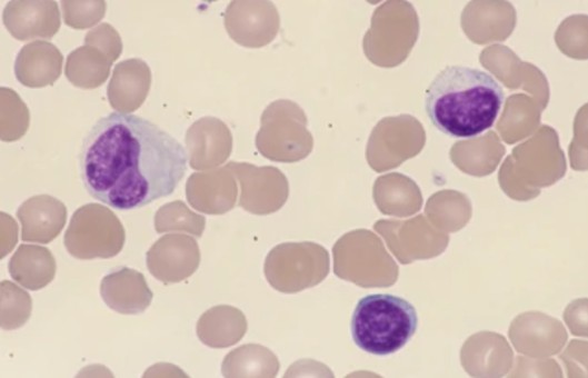
The preparation of DNA from human peripheral blood leukocytes, also known as white blood cells, is a common procedure in molecular biology and genetic research. To prepare leukocyte DNA from whole blood, the cell membranes of erythrocytes and leukocytes in the blood can be ruptured directly with the nonionic decontaminant Triton X-100, releasing hemoglobin and nuclei, and the osmotic pressure inside and outside the nuclear membrane is maintained by including a certain concentration of sucrose in the reaction system to prevent rupture of the nuclear membrane, and the nuclei of leukocytes can be obtained by centrifugation and other means. Adding SDS to the nuclear suspension disrupts the nuclear membrane and dissociates the DNA from the nucleoprotein, and leukocyte DNA can be obtained by conventional DNA preparation procedures such as Proteinase K and phenol/chloroform extraction.
METHODS
- Take 2-5 ml of peripheral blood, add 9 times the volume of pre-cooled solution I, shake for about 5-10 minutes until the solution is transparent, and centrifuge at 4°C 3000 × g for 10 minutes.
- Discard the supernatant, precipitate with 0.9% NaCl, wash 3000 × g, and centrifuge for 10 minutes.
- Discard the supernatant, add 5 ml of pre-cooled solution II suspension nuclear precipitation, add SDS to the final concentration of 0.5%, and add protease K to the final concentration of 200 μg/ml, mix well, and keep in a water bath at 65°C for 5 hours.
- Add 1/2 volume TE (pH 8.0), saturate, mix, rotate at room temperature for 3 minutes, add 1/2 volume chloroform/isoamyl alcohol, reverse the plastic tube, gently and fully mix, centrifuge at 600 × g room temperature for 3 minutes, the solution is divided into two phases, use a large plastic suction head to absorb the upper liquid in a clean centrifuge tube, and repeatedly extract until the interface is clean.
- Remove the upper liquid, extract it once with equal volume chloroform/isoamyl alcohol, and centrifuge at 600×g at room temperature for 3 minutes.
- Take the upper water phase, add 1/3 volume of 10mol/L NH4AC and 2 times the volume of pre-cooled anhydrous ethanol, and mix well to precipitate fibrous DNA from the solution.
- Use a large plastic suction head to suck out the filamped sediment, wash it once with 70% ethanol, drain it, dissolve it in an appropriate amount of 1 × TE (pH 8.0), and store it at 4°C for later use.
- Electrophoresis identification (see Restriction enzymes of plasmid DNA and electrophoresis analysis).
Creative Bioarray Relevant Recommendations
- Creative Bioarray is dedicated to providing high-quality peripheral blood mononuclear cells (PBMCs) for various biomedical and research applications. We aim to advance scientific and medical progress by supplying premium PBMCs processed using cutting-edge isolation and purification techniques.
- We provide a series of nucleic acid extraction kits for a variety of samples to help our customers accelerate their research. Beyond that, we also offer extraction services of nucleic acid from a wide range of starting materials.
| Cat. No. | Product Name |
| CNEK-D1791H | QualiNuclei® Genomic DNA Extraction Kit |
| CNEK-D1792H | QualiNuclei® Cells and Tissue DNA Extraction Kit |
| CNEK-D1793H | QualiNuclei® Cells and Tissue DNA Extraction Micro Kit |
| CNEK-D1794H | QualiNuclei® Cells and Tissue DNA Extraction Kit (Magnetic Bead) |
| CNEK-D1795H | QualiNuclei® Cells and Tissue DNA Extraction Kit (96-Well) |
| CNEK-D1798H | QualiNuclei® FFPE DNA Extraction Kit |
NOTES
- Proper collection techniques and sample handling are essential to prevent DNA degradation and maintain sample integrity. The use of EDTA-containing tubes for anticoagulation and immediate processing of the blood sample is crucial to avoid cell lysis and DNA damage.
- Gentle and thorough cell lysis is crucial to release intact genomic DNA and prevent shearing or degradation of the DNA molecules. The choice of lysis buffer and proteinase K concentration should be optimized to ensure maximum DNA yield and purity.
- Precipitation and purification steps require attention to detail to ensure the removal of contaminants and salts that can affect the quality of the DNA. Avoiding DNA over-drying and ensuring complete resuspension of the DNA pellet in an appropriate buffer are key considerations.