Animal Models of Necrotizing Enterocolitis
Seminars In Perinatology. 2023 Feb; 47(1): 151695.
Authors: Lopez CM, Sampah MES, Duess JW, Ishiyama A, Ahmad R, Sodhi CP, Hackam DJ
INTRODUCTION
Necrotizing enterocolitis (NEC) is the leading cause of death and disability from gastrointestinal disease in premature infants. The mortality of patients with NEC is approximately 30%, a figure that has not changed in many decades, reflecting the need for a greater understanding of its pathogenesis. Progress towards understanding the cellular and molecular mechanisms underlying NEC requires studying highly translational animal models. Such animal models must mimic the biology and physiology of premature infants while allowing for safe experimental manipulation of environmental and microbial factors that are thought to be associated with the risk and severity of NEC.
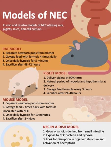 Fig. 1 Experimental models to study the pathogenesis of necrotizing small bowel colitis.
Mouse Models of NEC
- Mouse models are some of the earliest and most frequently utilized models to study NEC. In general terms, as with other diseases, mouse models offer the opportunity for genetic modification to test disease pathogenesis and have the advantage of being relatively inexpensive to purchase and house compared with other animals.
- While individual models vary somewhat, the typical mouse models are performed on mice within the first week or so of life and utilize a combination of gavage with infant formula, brief exposure to hypoxia and/or hypothermia, and the addition of either bacterial lipopolysaccharide or bacteria obtained from human infants with clinical NEC.
- Several approaches to validating the mouse and human NEC models have been performed. First, the newborn mouse shares features consistent with a 28-week-old infant, i.e., minimal subcutaneous fat, eyes closed most of the time, poor thermoregulation, and immature gut motility. Second, the newborn mouse with NEC exhibits a pattern of gene expression in component immune cells within the small intestine that resembles the human infant with NEC. Further, the intestinal microbiome seen in mice with NEC parallels that in humans. Most importantly, biochemical and genetic pathways that play causative roles in mouse NEC are also observed in human disease. Among these are pathways involving TLR4, IgA, EGF, and HMGB1.
- The complexity of the mouse model also gives rise to its inherent disadvantages: mouse models of NEC require regular feeding and a team of investigators trained to administer formula by gavage to tiny mice. The model typically lasts 4 or 5 days, upon which most mice will display the patchy intestinal edema, pneumatosis intestinalis, and necrosis that characterizes human NEC.
Rat Models of NEC
- Rats were the first animals used to create an experimental model of NEC. The introduction of bacteria into the formula led to NEC, thus revealing a role for bacterial colonization in NEC development. Additional laboratories have made further adaptations to the rat NEC model to study the effects of colonization with different species of bacteria found in the stool of infants with necrotizing enterocolitis, including Cronobacter sakazakii, Klebiella, and Clostridia. LPS has also been used to increase the extent of intestinal injury in the rat model.
- The advantages of using rats in NEC research include their low cost and relative tolerance of the model, while disadvantages center primarily on the inability to manipulate the genome to interrogate specific pathways involved in disease development. Rat models of NEC may have essential roles in pharmacokinetics studies and safety and efficacy studies of potential NEC therapies.
Piglet Models of NEC
- Piglets offer an attractive model for studying NEC because of their anatomical, physiological, and developmental similarities to the gastrointestinal tract of humans. The premature piglet model, in particular, offers specific advantages in that the model is performed on animals weighing between 1000-1300 g, which approximates the size of the human infant with NEC.
- NEC is induced by feeding 20 mL/kg of formula supplemented with enteric bacteria obtained from infants with surgical NEC five times daily. Piglets are housed in an incubator for the duration of the model and euthanized with intracardiac potassium chloride injection following anesthesia with ketamine. The piglet NEC model has been used to understand early NEC detection methods, potential therapies, and preventative strategies.
Creative Bioarray Relevant Recommendations
- Creative Bioarray provides validated and custom in vivo disease modeling and services. Our comprehensive animal-based disease models of different organ systems allow our clients to choose the most appropriate models to test promising compounds and optimize lead candidates.
| Service Types | Description |
| Oncology Models | Creative Bioarray provides a comprehensive range of oncology models, such as cell line-derived xenograft (CDX) models, patient-derived xenograft (PDX) models, syngeneic and orthotopic models, gene-edited animal models, chemically induced animal models. |
| Metabolic Disease Models | Animal models of metabolic diseases provide an opportunity to examine correlations among different metabolic parameters. Creative Bioarray provides professional drug testing services using these models. |
| Urology Disease Models | Creative Bioarray has successfully developed 3 urology disease models to help with drug development. The application of urology animal models helps increase our understanding of urology diseases and provides new approaches to improve the diagnosis and treatment of kidney injury. |
| Inflammation and Autoimmune Disease Models | We provide several validated models of inflammatory and autoimmune diseases, including arthritis, psoriasis, colitis, and immune disease (CIA, DTH, PCA, EAE). Each disease type has a different model that provides different disease parameters for your candidate compounds. |
- Our streamlined process can offer timely and dependable mouse model and PDX analysis (FISH) results, giving clients the flexibility to focus on other research priorities.
RELATED PRODUCTS & SERVICES
Reference
- Lopez CM, et al. (2023). "Models of necrotizing enterocolitis." Semin Perinatol. 47 (1), 151695.