CFSE Cell Proliferation Assay
Introduction
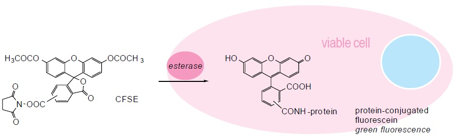
Amine-reactive dye CFSE (carboxyfluorescein diacetate, succinimidyl ester, also known as CFDA-SE) is widely used in cell proliferation and in vivo cell tracking. CFSE is non-fluorescent until it enters viable cells, where it is hydrolyzed by cytoplasmic esterase enzymes to release fluorescent amine-reactive dyes. These dyes then covalently react with the amine groups on intracellular proteins to form fluorescent conjugates that are retained in the cells. Moreover, due to this stable linkage, once the dyes are incorporated into the cells, they do not transfer to adjacent cells.
CFSE was originally developed to track lymphocyte migration. Subsequent studies revealed that this dye can be used to monitor cell proliferation both in vitro and in vivo, as CFSE fluorescence in progeny cells was gradually halved as the cells division.
Features
- Staining does not adversely affect cell health
- Stained cells retain long-term signal stability
- Bright and single-peak dyeing enables visualization of multiple generations
Materials
- CFSE
- Anhydrous DMSO
- PBS or other suitable buffer
- Aldehyde-containing fixative
CFSE Stock Solution
- Dissolve 25 mg of CFSE in 8.96 mL of anhydrous DMSO for a final stock solution of 5 mM. Vortex briefly to mix.
- To prepare the working solution, dilute the stock solution to a final working concentration in PBS or other non-amine containing buffer just before use.
Note: 1) CFSE dye reacts with amine group and should not be used with amine-containing buffers such as Tris-based buffers or with poly-lysine coated culture vessels and slides. 2) CFSE will react with aqueous solution so it is critical that such exposure be avoided during storage.
Method to Label Cells in Suspension
- Centrifuge and aspirate the supernatant to obtain cell pellet.
- Resuspend the cells gently in pre-warmed (37°C) PBS containing CFSE working solution at the appropriate concentration (1-10 μM).
- Incubate the cells for 20 minutes at 37°C to label the cells.
- Pellet the labeled cells by centrifugation and resuspend in fresh pre-warmed culture medium.
- Incubate the cells for another 20 minutes at 37°C to ensure sufficient hydrolysis of CFSE.
- For microplate quantitation of viable cells, proceed the next step. For flow cytometry analysis of cell division, proceed with cell stimulation, incubation, and analysis.
- Wash the cells in PBS or other similar buffer.
- Transfer cells to multiwell plate and measure fluorescence by microplate reader, or mount cells on a slide and analyze by fluorescence microscopy. For cell division tracking, analyze by flow cytometry.
Method to Label Adherent Cells
- Grow cells to desired density on coverslips or chamber slides with the appropriate culture medium.
- Remove the medium and add pre-warmed (37°C) PBS containing CFSE working solution at the appropriate concentration (1-10 μM).
Note: Use sufficient working solution to completely submerge the cells. - Incubate the cells for 20 minutes at 37°C to label the cells.
- Replace the labeling solution with fresh pre-warmed cell culture medium.
- Incubate for at least 20 minutes at 37°C to ensure sufficient hydrolysis of CFSE.
- For microplate quantitation or fluorescence microscopy, proceed to the next step. For flow cytometry tracking of cell division, proceed with cell stimulation, incubation, or analysis.
- Wash the cells in PBS or other similar buffer.
- Analyze by fluorescent microplate reader or fluorescence microscopy. For cell division tracking, detach cells from the substrate by trypsinization or other cell dissociation method and analyze by flow cytometry.
Fixation and Permeabilization (Optional)
- Before fixation, wash and resuspend the cells with PBS or other suitable buffer.
- Fix the cells for 15-20 minutes at room temperature using an aldehyde-containing fixative, protected from light.
- Wash the cells with PBS.
- If needed, permeabilize the cells by using any appropriate protocol such as incubation in ice-cold acetone for 10 minutes.
- Following permeabilization, wash the cells with PBS.
- Resuspend the cells in PBS prior to analysis.
Combining with Other Fluorescent Markers (Optional)
- Resuspend the cells in PBS for the subsequent staining applications.
- Apply stains for immunophenotyping, DNA content, apoptosis, or other markers as recommended for each stain.
Troubleshooting
| Problem | Possible Causes | Recommended Solutions |
| Low signal of CFSE | Cells not healthy | Use only healthy cells |
| Cells not well labeled by CFSE | Titrate CFSE to get an optimal staining concentration | |
| Cell death | CFSE concentration is too high, resulting in cytotoxicity | Titrate CFSE to get an optimal staining concentration |
| Experimental compound is resulting in cytotoxicity | Lower concentration of the experimental compound | |
| Broad peak of CFSE in unstimulated controls | Poor mixing of CFSE reagent with cells | Mix cells with CFSE immediately and well upon addition |
| Multiple cell types with different proliferative capacity in the well | Label cells with fluorochrome-conjugated antibodies to select specific populations |
References
- Lyons A. B. Analysing cell division in vivo and in vitro using flow cytometric measurement of CFSE dye dilution. J Immunol Methods, 2000, 243: 147-154.
- Parish C. R. et al.; Use of the intracellular fluorescent dye CFSE to monitor lymphocyte migration and proliferation. Current Protocols in Immunology, 2009, 49: 1-13.
Cell Services:
Cell Line Testing and Assays: