Visualization of Inflammatory Gene Products in Neuronal Cells by RNAscope and IHC
Frontier in Molecular Neuroscience. 2023 Aug 17; 16: 1225847.
Authors: Ball JB, McNulty CJ, Green-Fulgham SM, Dragavon JM, Correia Rocha IR, Finch MR, Prévost ED, Siddique II, Woodall BJ, Watkins LR, Baratta MV, Root DH.
INTRODUCTION
A challenge for central nervous system (CNS) tissue analysis in neuroscience research has been the difficulty to co-detect and colocalize gene and protein expression in the same tissue. Given the importance of identifying gene expression relative to proteins of interest, for example, cell-type specific markers, we aimed to develop a protocol to optimize their codetection. RNAscope fluorescent in situ hybridization (FISH) combined with immunohistochemistry (IHC) in fixed (CNS) tissue sections allows for reliable quantification of gene transcripts of interest within IHC-labeled cells.
METHODS
- Male Sprague-Dawley rats (n = 6; Inotiv, Indianapolis; 10 weeks of age at arrival) were housed in pairs in an AAALAC-approved animal facility on a 12-h light/dark cycle. After acclimation, all six rats received unilateral chronic constriction injury (CCI). Tissues were collected 7 days after CCI.
- At the time of tissue collection, rats were given a lethal dose of sodium pentobarbital. Lumbar spinal cords were isolated and post-fixed in 4% paraformaldehyde for 4 h at 4°C. The length of post-fixation time may need adjustment depending on the research context. Spinal cords were cryoprotected with sucrose solutions (15%, 20%, and 30% in PB) over sequential days (24 h per solution) at 4°C. Frozen sections of 14 μm thickness were cut in a cryostat at -18°C and collected onto slides.
- The slide-mounted tissue sections were prepared for RNAscope probe hybridization through a series of steps including dehydration, peroxide blocking, and target retrieval blocking. The next day, slides were incubated in RNAscope amplifier components and tagged with a fluorescent dye. Immediately after fluorescent RNA labeling, tissue sections were incubated overnight at 4°C in primary antibodies in solution with do-detection diluent. After overnight incubation, the tissue was gently washed in DI water and then incubated with secondary antibody solutions for 2 h. Finally, the slides were washed in DI water and coverslipped with mounting medium.
- Images are acquired using a laser-scanning confocal microscope. A Z stack image was used to show the stain in detail with complete cell morphology. The images acquired for analysis were 2D single confocal Z slices from the central optical plane of the stained tissue. Standard image analyses were undertaken to colocalize the RNAscope puncta and the labeled cell bodies using Imaris software. Briefly, RNAscope puncta above a brightness threshold were marked. The spatial position (X and Y values) for each marked puncta was entered into a database. The image channels for each IHC stain were analyzed independently.
- Browse our recommendations
| Product/Service Types | Description |
| Sample Collection Service | Creative Bioarray provides the pharmaceutical and biotech industry with sample collection and analysis services to researchers worldwide. Our capabilities include collecting patient samples, plasma, whole blood, serum, urine, saliva, tissue, and other biological specimens on request. |
| In Situ Hybridization (ISH) & RNAscope Service | Creative Bioarray offers Custom RNA ISH and ACD Bio's RNAscope service. We have extensive experience in the quantification of RNA expression on a cell-by-cell basis across a whole tissue section or within defined regions of interest. |
| Immunohistochemistry (IHC), Immunofluorescence (IF) Service | By providing high-quality immunohistochemistry (IHC) and immunofluorescence (IF) services to customers for many years, Creative Bioarray will give you the best and most comprehensive service in regular and customized immunohistochemistry and immunofluorescence services. |
| Custom Probes | Creative Bioarray is pleased to announce the introduction of custom-labeled ISH Probes for a range of molecular and cytogenetic applications. |
| IHC ISH Reagents | We are dedicated to providing high-quality reagents for IHC and ISH applications. Our comprehensive product portfolio includes a diverse range of reagents optimized for precision, sensitivity, and reliability in detecting and localizing specific proteins and nucleic acids within tissue samples. |
RESULTS
- Nerve injury (CCI) significantly increased laminae IV/V NLRP3 mRNA expression in the ipsilateral compared to the contralateral dorsal horn (p = 0.0053). Within IBA1+ regions of laminae IV/V, NLRP3 mRNA was increased ipsilaterally compared to contralateral (p < 0.0001), as was the density of NLRP3 mRNA contained within modeled microglia (p < 0.0110). The elevated mRNA density in modeled microglia indicates that the heightened NLRP3 mRNA expression is due to an increase in per-cell transcription, not merely from additional microglia proliferation or recruitment. CCI did not significantly increase either NLRP3 mRNA within NeuN+ regions (p = 0.5952), or the density of NLRP3 within NeuN+ regions of laminae IV/V compared to the contralateral site in dorsal horn (p = 0.0847).
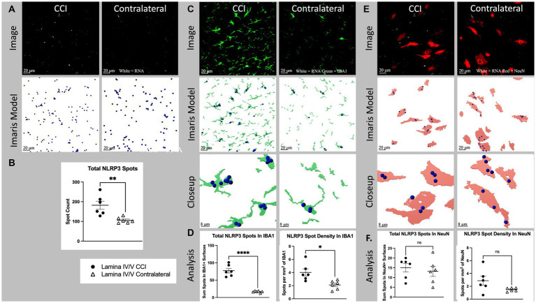 Fig. 1 Cell specific NLRP3 RNA expression in lamina IV/V lumbar (L3-6) dorsal horn ipsilateral to CCI vs. contralateral control.
- Nerve injury (CCI) significantly increased laminae IV/V IL-1b mRNA expression in ipsilateral compared to contralateral dorsal horn (p = 0.0364). Within IBA1+ regions of laminae IV/V, IL-1 mRNA was increased ipsilaterally compared to contralateral (p = 0.0011), as was the density of IL-1 (p = 0.0065). The increased mRNA density suggests that some of the difference in expression of IL-1 mRNA is due to increased transcription per cell, rather than being accounted for by proliferation and/or recruitment of additional microglia. CCI did not significantly increase either IL-1 mRNA within NeuN+ regions (p = 0.6211), or the density of IL-1 within NeuN+ regions of laminae IV/V compared to the contralateral site in dorsal horn (p = 0.3094).
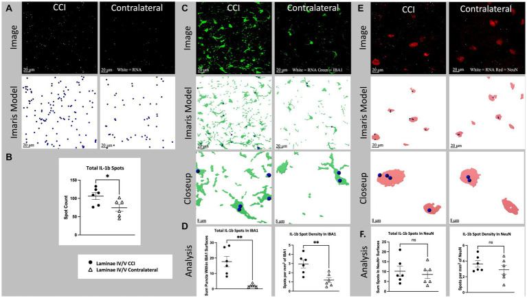 Fig. 2 Cell specific IL-1b RNA expression in lamina IV/V lumbar (L3-6) dorsal horn ipsilateral to CCI vs. contralateral control.
SUMMARY
This paper describes a new method for simultaneous visualization of FISH and IHC in thicker (14-μm), fixed tissue samples, using spinal cord sections. This method’s effectiveness is shown by the cell-type-specific quantification of two genes, namely the proinflammatory cytokine interleukin-1beta (IL-1b) and the inflammasome NLR family pyrin domain containing 3 (NLRP3).
RELATED PRODUCTS & SERVICES
Reference
- Ball JB, et al. (2023). "Combining RNAscope and immunohistochemistry to visualize inflammatory gene products in neurons and microglia." Front Mol Neurosci. 16: 1225847.