
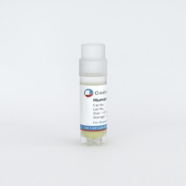
Human Nasal Epithelial Cells (HNECs)
Cat.No.: CSC-C1676
Species: Human
Source: Nose
Cell Type: Epithelial Cell
- Specification
- Background
- Scientific Data
- Q & A
- Customer Review
Human nasal epithelial cells (HNECs) are primary epithelial cells lining the nasal cavity. They can be obtained via minimally invasive nasal brushing or biopsy, providing an ethically favorable and repeatable source of donor-specific airway epithelium. When expanded and cultured at the air-liquid interface (ALI), HNECs differentiate into a pseudostratified mucociliary epithelium comprising basal, ciliated, goblet, and secretory cells. This model recapitulates key features of the human airway barrier, including tight junctions, mucus production, coordinated ciliary beating, ion transport (e.g., CFTR/ENaC), and innate immune signaling. Functional readouts include transepithelial electrical resistance (TEER), ciliary beat frequency, mucus secretion, and cytokine release.
Compared with immortalized cell lines, HNECs retain donor genetic and epigenetic backgrounds, native receptor expression, and functional responses, making them highly translatable for disease modeling and drug discovery. They are widely used to study cystic fibrosis, primary ciliary dyskinesia, chronic rhinosinusitis, allergic rhinitis, asthma, and respiratory viral infections such as SARS-CoV-2, influenza, and RSV. Their accessibility also enables longitudinal sampling and personalized medicine approaches, while donor diversity supports population-relevant pharmacology and toxicity testing.
HNECs offer several practical advantages: non-invasive collection, expandability, cryopreservation, and compatibility with ALI, microphysiological systems, and high-content imaging. They serve as a physiologically relevant surrogate for upper airway biology and, in many contexts, a useful proxy for lower airway epithelial function, though tissue-specific differences should be considered.
Limited Response of Primary Nasal Epithelial Cells to Bordetella Pertussis Infection
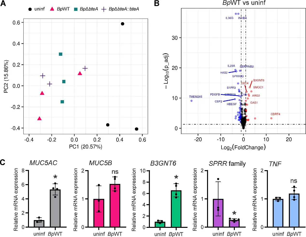
Here, Zmuda, Martin used primary human nasal epithelial cells (hNECs) cultured at the air-liquid interface and investigated their interaction with Bordetella pertussis B1917, focusing on the role of the type III secretion system effector protein BteA. In this model, which resembles the epithelial cells of nasal epithelium in vivo, B. pertussis B1917 localized predominantly in the overlying mucus and scarcely colonized the cell cilia. The colonization led to a gradual decline in epithelial barrier function, as shown by measurements of transepithelial electrical resistance (TEER) and staining of the tight junction protein zonula occludens 1. The decrease in TEER occurred independently of the cytotoxic effector protein BteA. Transcriptomic and proteomic analyses of hNECs showed only moderate changes following infection, primarily characterized by increased mucus production, including upregulation of mucin MUC5AC. No profound response to BteA was detected. Furthermore, the infection did not induce production of inflammatory cytokines, suggesting that B. pertussis B1917 evades recognition by hNECs in this model system. These results suggest that the mucus may serve as a niche that allows B. pertussis B1917 to minimize epithelial recognition and damage. The lack of a robust immune response further indicates that additional components of the nasal mucosa, such as innate immune cells, are likely required to initiate an effective host defense.
Ask a Question
Write your own review
Description: Human Thymic Epithelial Cells are isolated from human normal thymus. Poly-L-lysine-coated flasks are required for cell culture. Each vial contains at least 5x10^5 cells per mL.
Description: Mesothelial cells play pivotal roles in ovarian cancer metastasis, peritoneal dialysis, surgical adhesions, inflammatory response, and metabolic disease. These specialized epithelial cells form a ...
Description: Human Thymic Fibroblasts are isolated from human healthy thymus. HTyF are cryopreserved at passage one and delivered frozen. They are guaranteed to further expand for 15 population doublings ...
Description: Normal primary adult melanocytes are a vital research tool for a variety of applications from malignant melanoma research to pigmentation studies as well as for cosmetics and skin biology programs. ...
Description: Primary Human Fibroblast Cells are pooled primary isolates from 250 individual neonatal donor foreskins.These cells were originated using Complete Serum-Free Medium Kit With SuperFuel™, are ...
Description: Creative Bioarray provides Human Erythrocytes (RBCs) in a 10% suspension of phosphate-buffered saline (PBS).