
Human Thymic Epithelial Cells
Cat.No.: CSC-C9392W
Species: Human
Source: Thymus
Morphology: Polygonal
Cell Type: Epithelial Cell
- Specification
- Background
- Scientific Data
- Q & A
- Customer Review
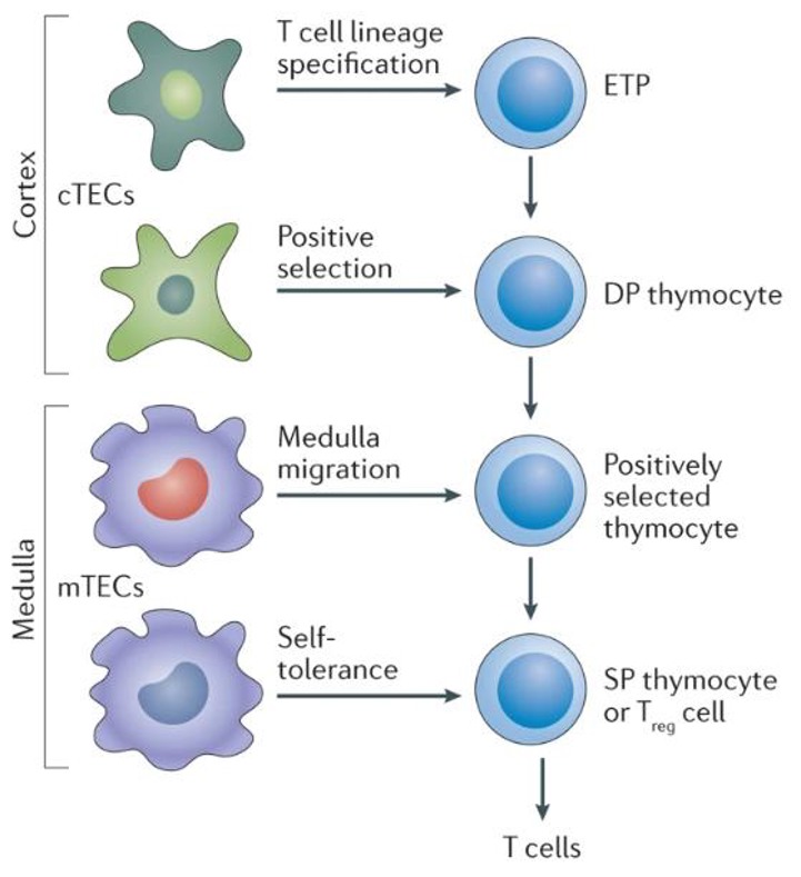
Human thymic epithelial cells (hTEC) form the stromal backbone of the thymus and drive T‑cell development. Two functionally distinct cortical (cTEC) and medullary (mTEC) cell subsets make up the thymic epithelial cell compartment. cTECs in the cortex express IL‑7, DLL4 and β5t and are involved in the positive selection of double‑positive thymocytes. In contrast, mTECs in the medulla express the autoimmune regulator (AIRE) and a wide array of self‑antigens, and are responsible for negative selection and central tolerance. Immortalized hTEC lines such as MTC, MITE, MITC and HTEC are derived by transduction with the SV40 large-T antigen, display an adherent polygonal morphology, express cytokeratins, UEA‑1, Claudin‑4 and MHC‑II and can be propagated in serum‑supplemented media.
hTEC are known to secrete cytokines (IL‑1, IL‑6 and TNF‑α) and chemokines, thereby programming the thymic microenvironment. They are used extensively to model in vitro T‑cell development, perform CRISPR‑mediated gene studies, screen immunomodulatory small molecules and drugs, and are a platform for studying thymic regeneration. Recently, the use of iPSC‑derived thymic epithelial precursors, to recapitulate cortical‑medullary architecture in culture has led to promise in cell‑based therapies to treat thymic hypoplasia and age‑related immune senescence.
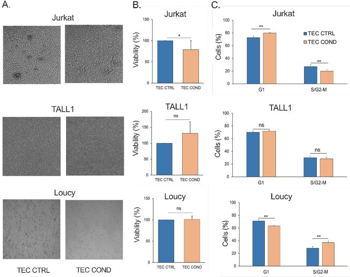
TEC-Conditioned Medium Provides a Microenvironment Influencing T-ALL Cell Viability
T-cell acute lymphoblastic leukemia (T-ALL) is a cancer marked by immature T-cell infiltration in the bone marrow. Aberrant NOTCH signaling, primarily due to NOTCH1 mutations and NOTCH3 overexpression, plays a critical role in T-ALL. However, the mechanisms by which pre-leukemic cells escape thymus retention and infiltrate the bone marrow remain unclear.
Patel et al. hypothesized that human thymic epithelial cells (hTECs) release soluble factors that could affect the viability and growth of T-ALL cell lines. They tested the effects of hTEC-conditioned medium (TEC COND) on Jurkat (NOTCH1-dependent), TALL1 (NOTCH3-dependent), and Loucy (NOTCH-independent) cells. After 24 hours, Jurkat cells showed reduced density in TEC COND compared to control medium (Fig. 1A), while TALL1 and Loucy cells did not change (Fig. 1A). Jurkat cells also had fewer live cells in TEC COND, indicating sensitivity to hTEC-derived factors (Fig. 1B). In contrast, TALL1 and Loucy cells were unaffected (Fig. 1B). They also examined how different culture conditions affected the cell cycle (G1, S/G2-M phases) of the three cell lines. Flow cytometry data (Fig. 1C) showed that TEC COND medium caused Jurkat cells to accumulate in G1, indicating a G1 arrest. TALL1 cells in TEC COND medium showed no significant changes in cell cycle distribution (Fig. 1C). In contrast, Loucy cells in TEC COND medium shifted from G1 to S/G2-M phase, with a slight increase in S phase cells (Fig. 1). Overall, hTECs can influence Jurkat cell viability and cell cycle through the extracellular microenvironment, which seems more conducive to TALL1 and Loucy cell growth.
Ask a Question
Write your own review
Description: Human Thyroid Microvascular Endothelial Cells are isolated from human thyroid tissue.

Description: Human Thyroid Epithelial Cells are isolated from normal human thyroid tissue.
Description: Human Thyrocyte Cells are isolated form normal human thyroid tissue. T25 flasks is required for cell adhension to the culture vessels. Grow cells in ECM-coated culture vessels with 5% CO2. Each vial ...

Description: HThF from Creative Bioarray are isolated from human thyroid tissue. HThF are cryopreserved at passage one and delivered frozen. Each vial contains >5 x 10^5 cells in 1 ml volume. HThF are ...