
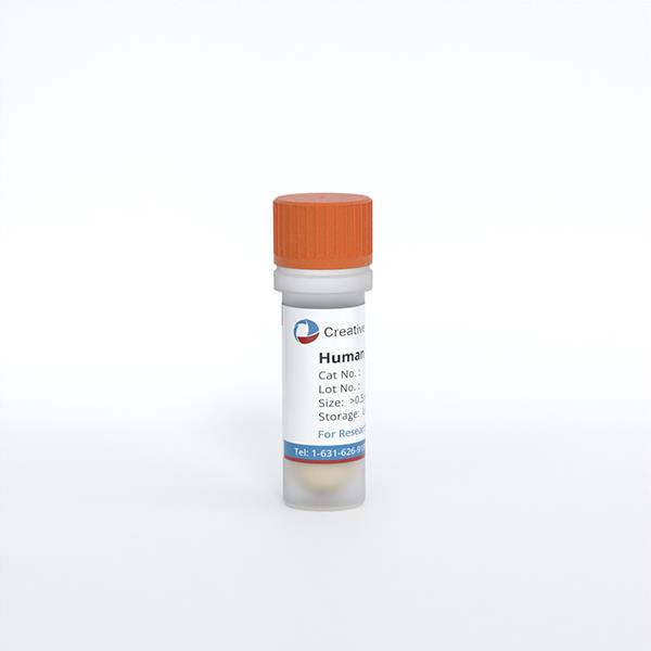
Human Hair Outer Root Sheath Cells (HHORSC)
Cat.No.: CSC-7773W
Species: Human
Source: Hair Follicle
- Specification
- Background
- Scientific Data
- Q & A
- Customer Review
Human Hair Outer Root Sheath Cells (HHORSC) are follicular epithelial keratinocytes derived from the outer root sheath of human hair follicles. This compartment contains distinct keratinocyte populations, including stem and progenitor cells associated with follicular renewal. HHORSC exhibit a characteristic epithelial keratin profile, including keratins 5, 14, and 17, providing a biologically relevant model for investigating human follicular epithelial behavior.
A key advantage of HHORSC is their accessibility and capacity for expansion. Viable outer root sheath keratinocytes can be isolated from appropriately plucked human hairs, allowing cell collection with minimal invasiveness. Under suitable culture conditions, these cells form colonies, undergo serial propagation, and can be cryopreserved. Their human origin and defined follicular source make them particularly useful for studying epithelial responses in a tissue-relevant context. Proliferative capacity varies with the follicular region sampled and the culture conditions employed.
HHORSC also display valuable differentiation plasticity. Under appropriate organotypic conditions, they can generate stratified epidermal tissue expressing terminal differentiation markers, supporting investigations of re-epithelialization, epidermal differentiation, and skin tissue engineering. Their responsiveness to dermal papilla-derived signals further enables studies of epithelial–mesenchymal communication, including the regulation of keratinocyte proliferation, migration, and follicular differentiation.
Activation of Mitochondrial Aldehyde Dehydrogenase 2 Promotes Hair Growth in Human Hair Follicles
Mitochondrial dysfunction-induced oxidative stress has been recognized as a crucial determinant of hair follicle (HF) biology. Aldehyde dehydrogenase 2 (ALDH2) mitigates oxidative stress by detoxifying acetaldehyde. To further investigate the role of ALDH2 in human HFs, Lee, Seunghee, et al. assessed its activity at the cellular level in ORSCs and DPCs using IF staining. The results showed that ALDH2 was primarily expressed in ORSCs.
To explore the impact of Alda-1 on HFs, they conducted an assessment of ALDH2 enzymatic activity in ORSCs. A significant increase was observed in ALDH2 activity in ORSCs after Alda-1 treatment (Fig. 1), indicating that Alda-1 functions as an ALDH2 activator in HFs, thereby modulating ALDH2 expression.
The cell permeating H2DCFDA is a widely employed probe for measuring ROS expression levels. To determine whether ALDH2 acts as a ROS scavenger in HFs, ROS levels were evaluated in ORSCs using H2DCFDA after Alda-1 treatment. The unstained group was also evaluated to confirm the effects of H2DCFDA on ROS expression levels. The results indicated that ROS levels were remarkably upregulated in each group after hydrogen peroxide (H2O2) treatment. After treatment with 5, 25, and 50 μM Alda-1, a dose-dependent reduction in ROS levels was observed. There was no difference in the cellular form, but the number of H2DCFDA-stained cells decreased in the presence of ROS (Fig. 2). ROS level reduction in ORSCs was further confirmed using flow cytometry. This observation highlights the potential role of ALDH2 activation in mitigating oxidative stress and modulating cellular redox homeostasis.
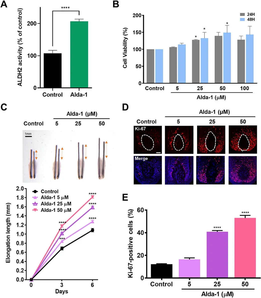
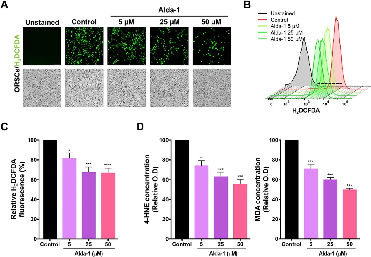 Fig. 2. ALDH2
activation induces ROS scavenging in ORSCs (Lee, Seunghee, et
al., 2024).
Ask a Question
Write your own review
Description: HHGMC from Creative Bioarray are isolated from germinal matrix of hair follicle bulbs. HHGMC are cryopreserved at passage one culture and delivered frozen. Each vial contains >5 x 10^5 cells in 1 ml ...
Description: The dermal papilla is a highly active group of cells. It is derived from the dermis mesenchyme, located at the base of the hair follicle. The dermal papilla is implicated in controlling the hair ...
Description: HHFK from Creative Bioarray are isolated from human hair follicles. HHFK are cryopreserved on primary culture and delivered frozen. Each vial contains >5 x 10^5 cells in 1 ml volume. HHFK are ...
Description: HHFIRSC from Creative Bioarray are isolated from human hair follicle inner root sheaths. HHFIRSC are cryopreserved at primary culture and delivered frozen. Each vial contains >5 x 10^5 cells in 1 ml ...