
Human Hair Dermal Papilla Cells
Cat.No.: CSC-C1490
Species: Human
Source: Hair Follicle
- Specification
- Background
- Scientific Data
- Q & A
- Customer Review
HHDPC are isolated from dermal papilla of human hair follicles. HHDPC are cryopreserved at passage one culture and delivered frozen. Each vial contains >5 x 10^5 cells in 1 ml volume. HHDPC are characterized by their mesenchymal cell morphology and immunofluorescent method with antibody to fibronectin and CD105. HHDPC are negative for HIV-1, HBV, HCV, mycoplasma, bacteria, yeast and fungi. HHDPC are guaranteed to further expand for 15 population doublings at the condition provided by Creative Bioarray.
Human Hair Dermal Papilla Cells (HHDPCs) are mesenchymal cells that have been harvested from the dermal papilla at the base of normal human scalp hair follicles (a niche of laminin- and type IV collagen-rich extracellular matrix that coordinates epithelial‑mesenchymal signaling during hair cycling). In vitro, they attach to plastic, exhibit a fibroblast‑like, spindle‑to‑polygonal shape, and show typical markers (alkaline phosphatase, α‑smooth‑muscle actin, versican and fibronectin) that differ them from dermal fibroblasts. These cells produce growth factors (EGF, IGF‑1, VEGF, KGF) and cytokines that influence anagen induction, sustain hair‑shaft production, and respond to androgen‑receptor signaling, which have made them the dominant model for the study of androgenic alopecia.
Functionally, in vitro cultured DPCs have the capacity to induce de‑novo hair follicle formation when transplanted in vivo, which forms the foundation for their widespread application in hair‑regeneration studies, as well as high‑throughput screening of hair‑growth promoting compounds. Recent developments include the integration of 3‑D scaffolds, extracellular‑matrix mimetics and immortalization approaches (hTERT, SV40 T/c‑Myc) to maintain inductivity for multiple passages, broadening their application in regenerative‑medicine platforms and mechanistic studies.
RSV Promotes Proliferation of Cultured HDPCs And Protects HDPCs Against Oxidative Stress
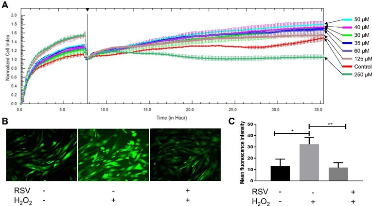
Oxidative damage has been found in various types of hair loss. As a polyphenolic phytoalexin, resveratrol (RSV) is known as an antioxidant, anti-inflammatory and anti-apoptotic agent. Thus, Zhang's team aims to examine the effects of RSV on hair growth.
DPC proliferation is crucial for hair growth and the hair cycle, as DPCs are key modulators in hair follicles. To assess the effect of RSV on hDPC proliferation, they added RSV at various concentrations to cultured cells. Lower doses of RSV (30-125 μM) accelerated hDPC proliferation (Fig. 1A), while a higher dose (250 μM) was cytotoxic. Therefore, they chose 50 μM RSV for subsequent ROS experiments. Since RSV is an antioxidant, they measured its ability to protect hDPCs from ROS using DCFH-DA as a probe. ROS was induced by H2O2. After 12 h, H2O2-treated cells showed significantly higher fluorescence intensity than controls (32.39 ± 5.97 vs 12.92 ± 6.32, *P = 0.0179). However, RSV pretreatment for 24 h prevented this increase (11.69 ± 4.40 vs 32.39 ± 5.97, **P = 0.0085) (Fig. 1B and C). Thus, RSV protects hDPCs from H2O2-induced oxidative damage.
Exosomes from Human Umbilical Cord Mesenchymal Stem Cells Promote the Growth of Human Hair Dermal Papilla Cells
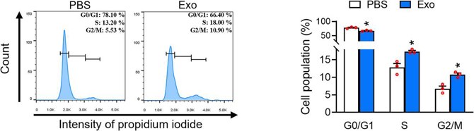
Human hair dermal papilla cells (HHDPCs) play a significant role in hair growth. Exosomes derived from umbilical cord mesenchymal stem cells (UC-MSCs) have proven highly valuable in skin repair. Chen's team explored the human umbilical cord mesenchymal stem cell-derived exosomes (UC-MSC-Es) on cell growth of HHDPCs.
The effect of UC-MSC-Es on HHDPC proliferation was tested. Fig 2 shows that UC-MSC-Es increased HHDPC proliferation in a concentration-dependent manner at 1 × 10¹⁰, 3 × 10¹⁰, and 1 × 10¹¹ particles/mL. Minoxidil (3 and 10 μM) served as a positive control. UC-MSC-Es had a significantly stronger effect than minoxidil (Fig. 2). To explore how UC-MSC-Es affect HHDPC proliferation, they treated HHDPCs with UC-MSC-Es at 1 × 10¹¹ particles/mL for 48 h. The treated cells had fewer cells in the G1 phase and more in the S and G2/M phases (Fig. 3). This suggests that UC-MSC-Es promote HHDPC proliferation by advancing the cell cycle to the S and G2/M phases.

Ask a Question
Write your own review
Description: HHFORSC from Creative Bioarray are isolated from human hair follicle outer root sheath. HHFORSC are cryopreserved at primary culture and delivered frozen. Each vial contains >5 x 10^5 cells in 1 ml ...
Description: HHFK from Creative Bioarray are isolated from human hair follicles. HHFK are cryopreserved on primary culture and delivered frozen. Each vial contains >5 x 10^5 cells in 1 ml volume. HHFK are ...

Description: HHGMC from Creative Bioarray are isolated from germinal matrix of hair follicle bulbs. HHGMC are cryopreserved at passage one culture and delivered frozen. Each vial contains >5 x 10^5 cells in 1 ml ...

Description: HHFIRSC from Creative Bioarray are isolated from human hair follicle inner root sheaths. HHFIRSC are cryopreserved at primary culture and delivered frozen. Each vial contains >5 x 10^5 cells in 1 ml ...