
Human Uterine Smooth Muscle Cells (HUSMCs)
Cat.No.: CSC-C4012X
Species: Human
Source: Uterus
Cell Type: Smooth Muscle Cell
- Specification
- Background
- Scientific Data
- Q & A
- Customer Review
Human Uterine Smooth Muscle Cells (HUSMCs) are isolated from the uterus tissue of healthy donors. The cells are cryopreserved at Passage 2 and delivered frozen. Each vial contains at least 0.5 *10^6 cells. The cells are negative for HIV-1, HBV, HCV, mycoplasma, bacteria, yeast and fungi. Human Uterine Smooth Muscle Cells are guaranteed for at least 15 population doublings under the conditions provided by Creative Bioarray. Repeated freezing and thawing of cells is not recommended.
Human uterine smooth muscle cells (HUSMCs) are primary myometrial cells isolated from human uterine tissue obtained during hysterectomy or biopsy. They provide a donor-specific, physiologically relevant in vitro model of uterine smooth muscle. Under optimized culture conditions, HUSMCs express canonical smooth muscle markers, including α-smooth muscle actin, desmin, calponin, SM22α, and smooth muscle myosin heavy chain, and retain key functional properties such as calcium signaling, agonist-induced contractility, and responsiveness to oxytocin, prostaglandins, estrogens, and progestins.
Compared with immortalized or animal-derived lines, HUSMCs better preserve native receptor expression, signaling pathways, and donor genetic background, making them highly translatable for studying uterine physiology and pathology. They are widely used to investigate myometrial contractility, preterm labor, dysmenorrhea, leiomyoma (fibroids), adenomyosis, and uterine fibrotic remodeling. Their hormone and inflammatory responsiveness also support drug discovery, including tocolytics, uterotonics, progesterone receptor modulators, and anti-fibrotic agents.
Practical advantages include expansion at low passage, cryopreservation, and compatibility with 2D and 3D cultures, collagen gel contraction assays, calcium imaging, electrophysiology, flow cytometry, ELISA, and transcriptomic profiling. HUSMCs can be matched with donor clinical data and compared with leiomyoma-derived cells, enabling personalized and mechanistic studies.
ASC-Derived Extracellular Vesicles Suppress Macrophage-Driven Inflammatory Amplification and Contractile Activation of Uterine Smooth Muscle Cells
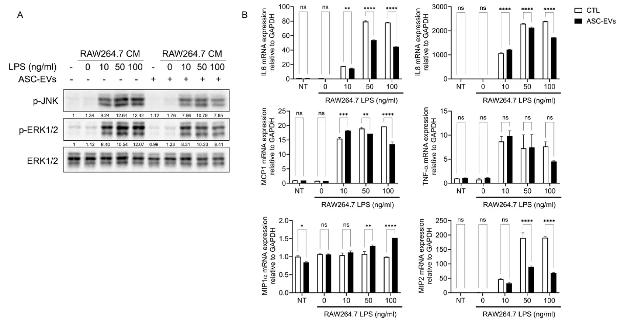
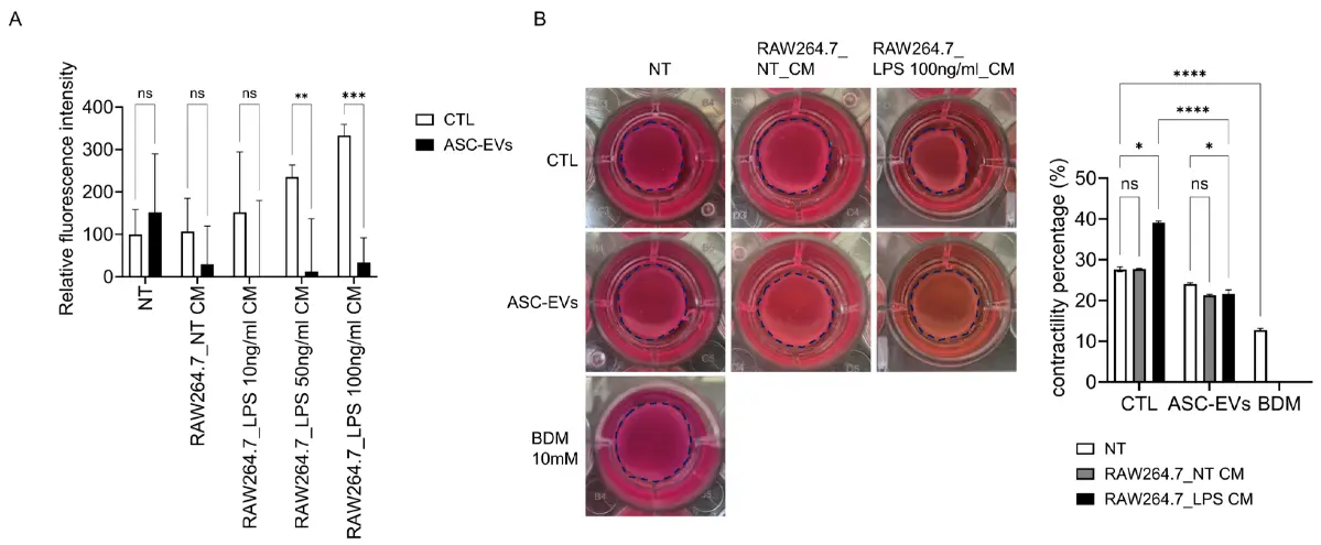
Preterm labor is a major cause of neonatal morbidity and mortality and is frequently driven by infection-associated inflammation that promotes premature uterine activation. In this study, we investigated the effects of adipose stem cell-derived extracellular vesicles (ASC-EVs) on macrophage-mediated inflammatory signaling in uterine smooth muscle cells (HUtSMCs). An in vitro model was established by treating HUtSMCs with conditioned media derived from LPS-stimulated RAW264.7 macrophages. Activation of signaling pathways was assessed by Western blotting and immunofluorescence, and functional responses were evaluated using calcium flux and collagen gel contraction assays.
Conditioned media from LPS-stimulated macrophages induced robust activation of MAPK (ERK1/2 and JNK) and NF-κB signaling, accompanied by IκB degradation and nuclear translocation of phosphorylated p65, whereas ASC-EVs pretreatment significantly attenuated these responses and reduced the expression of pro-inflammatory cytokines, including IL-6, IL-8, and MCP-1. Furthermore, macrophage-conditioned media enhanced intracellular calcium flux and contractile activity in HUtSMCs, both of which were suppressed by ASC-EVs. Inhibition of TLR4 signaling in macrophages reduced the inflammatory potency of conditioned media, indicating a key upstream role of macrophage TLR4 activation. Collectively, these findings demonstrate that ASC-EVs suppress macrophage-mediated inflammatory activation and downstream contractile responses, suggesting their potential as a cell-free therapeutic strategy for preventing inflammation-associated preterm labor.
Ask a Question
Write your own review
Description: Human Metastatic Ovarian Tumor -Associated Endothelial Cells are isolated from human ovarian metastases tissue developed from colon cancer. Human Metastatic Ovarian Tumor -Associated Endothelial ...
Description: HOF from Creative Bioarray are isolated from human ovary tissue. HOF are cryopreserved at primary culture and delivered frozen. Each vial contains >5 x 10^5 cells in 1 ml volume. HOF are ...
Description: Human Uterine Epithelial Cells are isolated from normal human uterine tissue.
Description: GFP Expressing Human Uterine Microvascular Endothelial Cells (GFP-HUMECs) provided by Creative Bioarray are puromycin-selected from Human Uterine Microvascular Endothelial Cells infected with ...
Description: HAEpiC from Creative Bioarray are isolated from human amniotic membranes. HAEpiC are cryopreserved at passage one and delivered frozen. Each vial contains >5 x 10^5 cells in 1 ml volume. HAEpiC are ...
Description: Human Ovarian Tumor-Associated Endothelial Cells from Creative Bioarray are isolated from human ovarian tumor tissue. Human Ovarian Tumor-Associated Endothelial Cells are grown in T25 tissue culture ...