
Rat Primary Tracheal Fibroblasts
Cat.No.: CSC-C4142X
Species: Rat
Source: Trachea
Cell Type: Fibroblast
- Specification
- Background
- Scientific Data
- Q & A
- Customer Review
Cells are negative for bacteria, yeast, fungi, and mycoplasma. Rat Primary Tracheal Fibroblasts are tested for expression of marker using the antibody of anti-FSP1/S100A4 by immunofluorescence staining and can be expanded by 2-4 passages at a split ratio of 1:2 under the cell culture conditions specified by Creative Bioarray. Repeated freezing and thawing of cells is not recommended.
Standard biochemical procedures performed with fibroblast cultures include the assays of cell to cell interaction, PCR, Western blotting, immunoprecipitation, immunofluorescent staining, immunofluorescent flow cytometry or generating cell derivatives for desired research applications.
Rat primary tracheal fibroblasts are isolated directly from the tracheal tissue of healthy donor rats, usually via enzymatic digestion and differential adhesion. These cells are a major stromal component of the airway wall, residing in the lamina propria and submucosa, where they synthesize and remodel extracellular matrix (ECM) components - notably collagens type I and III, fibronectin, and proteoglycans - thus providing structural integrity and biomechanical support to the trachea.
The principal advantage of rat primary tracheal fibroblasts is their physiologically relevant, in vivo‑like phenotype. Unlike immortalized cell lines, primary cells retain their native expression profiles, growth kinetics, and responsiveness to microenvironmental cues (e.g., TGF‑β, mechanical stretch, hypoxia) without the confounding genetic drift or transformation artefacts introduced by immortalization. This makes them an invaluable model for studying airway remodeling and fibrosis in respiratory diseases such as asthma, chronic obstructive pulmonary disease (COPD), and post‑intubation tracheal stenosis.
Additional advantages include: (i) they allow direct mechanistic investigation of fibroblast‑to‑myofibroblast differentiation (α‑SMA upregulation, stress fibre formation) - a key event in airway fibrosis; (ii) they can be co‑cultured with primary tracheal epithelial cells to reconstruct the epithelial‑mesenchymal trophic unit, enabling studies of crosstalk in inflammation and repair; (iii) they are amenable to pharmacological intervention, gene silencing, and mechanical loading studies; and (iv) they offer species relevance for pre‑clinical testing of anti‑fibrotic drugs before moving to larger animal models. Although primary tracheal fibroblasts have a limited proliferative lifespan, their phenotypic fidelity and physiological responsiveness remain the gold standard for ex vivo airway biology, particularly in the context of personalized respiratory medicine and toxicological evaluation of inhaled substances.
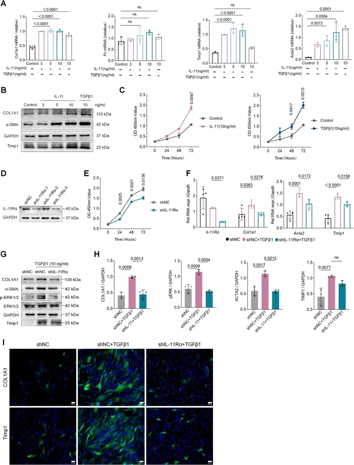
IL-11 Induces the Proliferation and Transformation of Rat Tracheal Fibroblasts into Myofibroblasts
Tracheal stenosis (TS) is a multifactorial and heterogeneous disease that can easily lead to respiratory failure and even death. Interleukin-11 (IL-11) has recently received increased attention as a fibrogenic factor, but its function in TS is uncertain.
To explore whether IL-11 is involved in profibrotic responses, we stimulated primary rat tracheal fibroblasts (PRTF) with an IL-11 concentration gradient. qPCR and WB indicated that IL-11 dose-dependently induced the expression of COL1A1, fibronectin (FN), α-SMA, and tissue inhibitor of metalloproteinases (TIMP1) in PRTF cells (Fig. 1A, B). In the CCK8 experiment, the OD values of the IL-11 and TGFβ1-treated PRTF cells were significantly higher than that of the control group after 72 h of incubation (Fig. 1C). These data indicate that IL-11 significantly promoted PRTF activation, proliferation, and secretion of extracellular matrix-associated proteins such as collagen type I, FN, α-SMA, and TIMP1. Therefore, we sought to understand whether inhibiting IL-11 signaling could block PRTF activation. After we transfected lentivirus in PRTF, the OD of cells in the shIL-11Rα group was significantly lower than in the shNC group, indicating that the IL-11Rα knockdown could inhibit cell proliferation (Fig. 1D, E). Then, we stimulated PRTF with TGFβ1 in the presence of shIL-11Rα. qPCR and WB showed that TGFβ1 significantly increased the mRNA and protein expression of α-SMA, TIMP1, and COL1A1 in cells (Fig. 1F-H). IF staining also indicated that COL1A1 and TIMP1 expressing positive cells were increased in the shNC+TGFβ1 group (Fig. 1I). However, compared with the shNC+TGFβ1 group, α-SMA, TIMP1 and COL1A1 expression were significantly reduced in the shIL-11Rα + TGFβ1 group (Fig. 1F-I). These studies demonstrate that IL-11 elicits profibrotic responses in PRTF and that TGFβ1-driven profibrotic effects partially depend on IL-11 activity.
Ask a Question
Write your own review
Description: Characterization: 1. Cell Activity; Recovery Viability ≥ 80%. 2. Cell Purity; - GFAP-positive cell percentage ≥ 70%. - β-tubulin III- or doublecortin- (Dcx-) positive cell percentage ≤ 10%. - ...

Description: Chondrocytes are the cells responsible for cartilage formation, and they are crucial for the process of endochondral ossification, which is useful for bone development. Ear chondrocytes represent the ...

Description: The thoracic aorta is located in the chest cavity and gives off arteries that branch to the esophagus, pericardium, lungs, and trachea. The thoracic aorta can be subdivided into the ascending aorta, ...

Description: Human Thymic Fibroblasts are isolated from human healthy thymus. HTyF are cryopreserved at passage one and delivered frozen. They are guaranteed to further expand for 15 population doublings ...
Description: Rat Bone Marrow Neutrophils are derived from the tibias and femurs of week-5 Sprague–Dawley Rats. Rat Bone Marrow Neutrophils are immediately cryo-preserved or fresh cells in suspension after ...
Description: Rat Bone Marrow Macrophages are derived from the tibias and femurs of 5 weeks old Sprague–Dawley Rats. Rat Bone Marrow Macrophages are grown in 100 mm treated tissue culture dish and incubated in ...