Porcine PB Mononuclear Cells
Cat.No.: CSC-C4421X
Species: Pig
Source: Peripheral Blood; Blood
Cell Type: Mononuclear Cell
- Specification
- Background
- Scientific Data
- Q & A
- Customer Review
Porcine peripheral blood mononuclear cells (PBMCs) are not technically considered a "cell line", but a primary cell population isolated from a pig blood donor. This distinction matters for both experimental design and interpretation of your data. PBMCs represent a mixed population of immune cells harvested through density gradient centrifugation (Ficoll-Paque for example) and are enriched in lymphocytes (T cells, B cells, NK cells) and monocytes.
Porcine PBMCs have strong utility as an intermediary between in vitro experiments and preclinical in vivo porcine models due to the physiological and immunological relevance of the pig. Immune cells are often leveraged to study human diseases and drug candidates. Porcine PBMCs can be used in studies involving immunology, virology, vaccine development. Common uses include: understanding host-pathogen interactions (African swine fever virus, influenza etc.), vaccine immunogenicity testing, cytotoxic T-cell function and antibody-mediated immunity, and immuno-oncology research to develop porcine tumor models or CAR therapies. PBMCs are advantageous over rodent models due to the closer translational match to human biology. As with any primary cells, they best represent the in vivo immune status of the donor but have limited life span in culture. They can be activated to live longer (mitogens such as ConA or PHA) or expanded for experimental use.
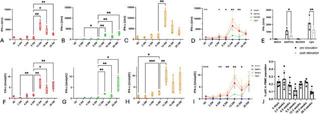
Age-dependent Differences in IFN-α Production by PBMC/pDC in Response to PRV-infected Cells or a TLR9 Agonist
Pseudorabies virus (PRV) causes severe disease with high mortality in young piglets but milder respiratory issues in older pigs. Claeys et al. report marked age-dependent differences in cytokine responses of primary porcine peripheral blood mononuclear cells (PBMCs) to PRV-infected cells.
Blood was collected from 5 piglets at 4 days to 28.5 weeks of age. PBMCs were isolated and incubated for 22 hours with mock-infected or PRV-infected (virulent Becker or attenuated Bartha strains) swine testicle cells. TLR9 stimulation with CpG ODN32 was included as control. IFN-α production was quantified in supernatants (Fig. 1A-D). Flow cytometry showed pDC percentages in PBMC fluctuated with age but without clear age-dependent trends (Fig. 1J).
The degradation of L-glutamine leads to the formation of ammonia, which is toxic to some cells.
Ask a Question
Average Rating: 5.0 | 1 Scientist has reviewed this product
Full knowledge
I am very grateful to this company for the full knowledge and technical support to accompany me in my scientific research.
02 Aug 2023
Ease of use
After sales services
Value for money
Write your own review
Description: Porcine Corneal Epithelial Cells from Creative Bioarray are isolated from corneal tissue of porcine. Porcine Corneal Epithelial Cells are grown in a T25 tissue culture flask pre-coated with ...
Description: Porcine Brain Vascular Fibroblasts from Creative Bioarray are isolated from brain tissue of porcine. Porcine Brain Vascular Fibroblasts are grown in T75 tissue culture flasks pre-coated with ...

Description: Porcine Thymus Endothelial Cells from Creative Bioarray are isolated from thymus tissue of porcine. Porcine Thymus Endothelial Cells are grown in T25 tissue culture flasks pre-coated with ...
Description: Porcine Kidney Endothelial Cells from Creative Bioarray are isolated from kidney tissue of porcine. Porcine Kidney Endothelial Cells are grown in T25 tissue culture flasks pre-coated with ...

Description: Pig bone marrow from Creative Bioarray is procured from the fresh femurs. Bone Marrow-CD34+ stem/progenitor cells are positively isolated using a direct immunomagnetic CD34 MicroBead labeling system.
Description: Porcine Primary Thymus Fibroblasts from Creative Bioarray are isolated from thymus tissue of porcine. Porcine Primary Thymus Fibroblasts are grown in T75 tissue culture flasks pre-coated with ...