
Human Thyroid Fibroblasts (HThF)
Cat.No.: CSC-7707W
Species: Human
Source: Thyroid
Cell Type: Fibroblast
- Specification
- Background
- Scientific Data
- Q & A
- Customer Review
Human Thyroid Fibroblasts (HThF) are human primary fibroblast cells derived from thyroid tissue. These cells have a spindle shaped morphology typical of fibroblasts and express various ECM proteins including fibronectin. Fibroblasts are mesenchymal derived cells that make up the connective tissue in our bodies. Within the thyroid gland fibroblasts are located in the interfollicular stroma and play roles in maintaining homeostasis as well as wound repair and ECM remodeling. HThF is an immortalized, healthy primary cell line that can easily be maintained in culture and allow for examination of thyroid specific cell types other than thyrocytes. Fibroblasts have been used extensively in research due to their high proliferation rates and ability to easily transfect and genetically modify these cells. In the thyroid gland fibroblasts have recently been shown to play dynamic roles within both physiological and disease states through communication with epithelial and immune cells.
Thyroid fibroblasts have begun to be used in modeling disease. For example, thyroid cancer cell lines can be co-cultured with thyroid fibroblasts to observe recruitment and activation of fibroblasts by cancer cells. Fibroblasts have also been used as a system to study hormone responsiveness. In one study, thyroid fibroblasts were used to determine alterations in expression of thyroid hormone responsive genes.
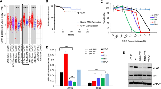
Overexpression of GPX4 and Targeting Ferroptosis in Thyroid Cancer Cells.
Metastatic papillary thyroid carcinoma (PTC) is associated with worse survival rates for patients. Metabolic processes such as the antioxidant glutathione (GSH) can modulate tumor progression. GPX4 is an enzyme that consumes GSH to regulate lipid peroxidation during oxidative stress; inhibition of GPX4 leads to ferroptosis. Sekher et al. aimed to investigate ferroptosis as a potential target for thyroid cancer treatment.
They analyzed TCGA database (N=501 PTC specimens) through TIMER 2.0 and found GPX4 is overexpressed in PTC tissue compared to benign tissue controls (Fig. 1A). GPX4 overexpression occurred in 45.1% of patients and was significantly associated with decreased 5-year overall survival (Fig. 1B). Thus, GPX4 overexpression is both prevalent and negatively associated with prognosis in patients with thyroid cancer.
Treatment with the GPX4 inhibitor RSL3 had differential effects on cell viability for different thyroid cancer cell lines. LC50 values were significantly decreased for RAS/BRAF mutant lines compared to human thyroid fibroblast controls (HThF). RSL3 treatment did not significantly decrease viability in TPC-1 cells that lack mutant forms of RAS/BRAF, illustrating toxicity selectively in RAS/BRAF-addicted cells (Fig. 1C).
RT-qPCR and Western blot confirmed expression of GPX4 and Transferrin Receptor 1 (TfR1) was heterogeneous across thyroid cancer cell lines. K1 cells overexpress GPX4 and TfR1 when compared to other cell lines MDA-T32, MDA-T68, TPC-1 and non-tumor controls (HThF) (Fig. 1D and E). GPX4/TFR1 expression heterogeneity in cell lines recapitulates patient data and highlights that GPX4/TFR1 may not be uniform markers of ferroptosis in vitro.
Ask a Question
Write your own review
Description: Human Thyroid Microvascular Endothelial Cells are isolated from human thyroid tissue.
Description: Human Thyroid Epithelial Cells are isolated from normal human thyroid tissue.

Description: Human Thyrocyte Cells are isolated form normal human thyroid tissue. T25 flasks is required for cell adhension to the culture vessels. Grow cells in ECM-coated culture vessels with 5% CO2. Each vial ...
Description: Human Thymic Epithelial Cells are isolated from human normal thymus. Poly-L-lysine-coated flasks are required for cell culture. Each vial contains at least 5x10^5 cells per mL.