

Human Amniotic Mesenchymal Stromal Cells (HAmMSC)
Cat.No.: CSC-7841W
Species: Human
Source: Placenta
Cell Type: Stromal Cell
- Specification
- Background
- Scientific Data
- Q & A
- Customer Review
Human Amniotic Mesenchymal Stromal Cells (HAmMSCs) are multipotent mesenchymal stromal cells isolated from the human amniotic membrane; a perinatal tissue discarded after delivery. Their collection is non-invasive to mother and infant and raises minimal ethical concerns. HAmMSCs express canonical MSC markers CD73, CD90, and CD105, while lacking CD34, CD45, and HLA-DR, and show low expression of costimulatory molecules together with immunomodulatory HLA-G.
Their key advantages are low immunogenicity and potent immunomodulation. HAmMSCs suppress T-cell and NK-cell activation, promote regulatory T cells, and shift macrophages toward anti-inflammatory phenotypes. They secrete trophic, anti-inflammatory, pro-angiogenic, and anti-fibrotic factors, supporting tissue repair and regeneration. Compared with bone marrow MSCs, HAmMSCs are abundant, proliferate rapidly, and can be expanded and cryopreserved for scalable banking. They retain multilineage differentiation potential (osteogenic, chondrogenic, adipogenic) under defined conditions and are generally considered low-risk for tumorigenicity.
Applications include regenerative medicine, wound healing, corneal repair, graft-versus-host disease, autoimmune and inflammatory disorders, and tissue engineering. They are also used in co-culture, immunomodulation, and drug-screening studies.
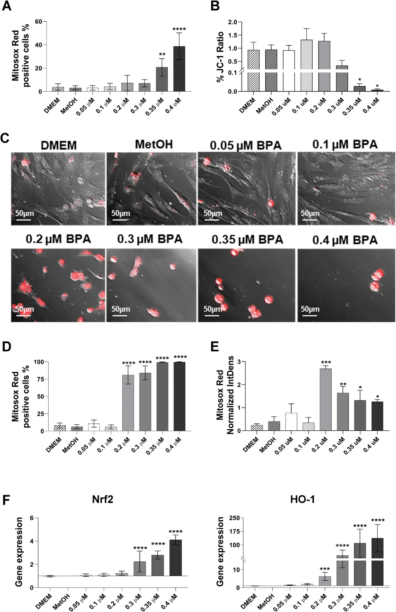
Bisphenol-A Disrupts Mitochondrial Functionality Leading to Senescence and Apoptosis in Human Amniotic Mesenchymal Stromal Cells
Among the degradation by-products of microplastics, Bisphenol-A (BPA) has emerged as a hazardous chemical, with potential toxicity at multisystemic level, particularly on the earliest stages of human development. Based on these findings, this study focuses on assessing the impact of BPA on properties and functions of mesenchymal stromal cells isolated from the amniotic membrane (hAMSC) of the human term placenta.
The results revealed how exposure to increasing concentrations of BPA compromise mitochondrial functionality in hAMSC, resulting in enhanced production of reactive oxygen species at mitochondrial level (mtROS). This, in turn, leads to the stabilization of p53, which triggers an increased expression of p21 and p27 encoding genes and an imbalance in the genetic expression of Bax and Bcl-2. Additionally, upregulated expression of cytokines and chemokines associated with the senescence-associated secretory phenotype (SASP) was observed. The increased oxidative stress, which plays a central role in BPA-mediated toxicity, can trigger the activation of the senescence pathways, or culminate in cell death, due to the overwhelming stress conditions. Therefore, these results provide novel insights into the mechanism of action of BPA and elucidates its impact on the functionality of hAMSC.
Ask a Question
Write your own review
Description: Human Placental Vascular Endothelial Cells are isolated from normal fetal placenta tissue. 25 flasks is required for cell adhension to the culture vessels. Grow cells in ECM-coated culture vessels ...
Description: Human Chorionic Mesenchymal Stem Cells (hCMesenchymal Stem Cells) are derived from extraembryonic mesoderm and are isolated from fresh chorion following mechanical and enzymatic removal of the ...
Description: HCMSC from Creative Bioarray are isolated from human chorionic membrane. HCMSC are cryopreserved at passage one culture and delivered frozen. Each vial contains >5 x 10^5 cells in 1 ml volume. HCMSC ...
Description: Human Placental Microvascular Pericytes (HPMPs) are isolated from normal human placental tissue. The cells are cryopreserved at passage 2 and delivered frozen. Each vial contains more than 0.5*10^6 ...
Description: Human Placental Epithelial Cells (HPECs) are isolated from the inner surface of the amnion. The cells are cryopreserved at passage 2 (P2) and delivered frozen. Each vial contains more than 0.5*10^6 ...
Description: Human Placental Microvascular Endothelial Cells are isolated from human placental tissue.