
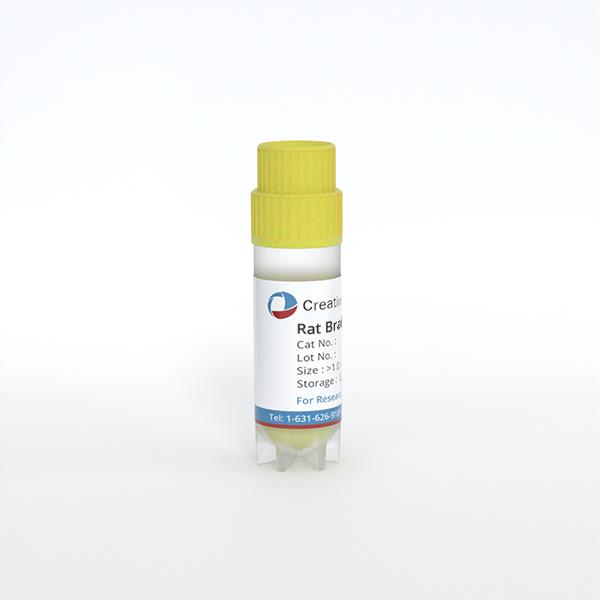
Rat Brain Cortex Neurons
Cat.No.: CSC-C8052L
Species: Rat
Source: Brain
Cell Type: Neuron
- Specification
- Background
- Scientific Data
- Q & A
- Customer Review
Rat Brain Cortex Neurons are primary neurons isolated from the rat cerebral cortex, providing a model for investigating cortical neuronal development, signaling, and network function. Under appropriate culture conditions, these cells establish neuronal polarity, extend axons and dendrites, and form functional synaptic connections. Their cortical origin and capacity for functional maturation make them particularly useful for linking neuronal morphology with electrophysiological activity.
A principal advantage is their ability to generate interconnected networks with measurable spontaneous electrical activity. As cultures mature, changes in action-potential firing, bursting, and coordinated network activity can be monitored using patch-clamp electrophysiology or multielectrode arrays. Complementary calcium imaging and immunocytochemistry enable assessment of intracellular signaling, neurite architecture, and synaptic organization. This combination supports investigation across molecular, cellular, and network levels within a controlled experimental system.
Rat cortical neuronal cultures also support studies of neurotransmitter receptor regulation and activity-dependent plasticity. Research has demonstrated maturation-associated changes in AMPA receptor expression and homeostatic regulation of glutamatergic and GABAergic synaptic components. These properties provide informative endpoints for examining how pharmacological interventions or environmental perturbations influence synaptic transmission and network stability. Neuron–glia co-culture formats further enable investigation of cellular interactions that support neuronal survival and function.
AKR1C1 Protects Against Intracerebral Hemorrhage by Suppressing Neuronal Cell Death
Intracerebral hemorrhage (ICH) is associated with the highest rates of mortality and residual disability. To date, effective treatments to delay or prevent ICH are still lacking. Multiple forms of neuronal cell death have been discovered following ICH, including apoptosis, necrosis, autophagy, and ferroptosis. Aldo-keto reductase family 1 member C1 (AKR1C1) has been identified to act as a protective factor in ferroptosis. However, whether AKR1C1 was involved in the development of ICH was unknown.
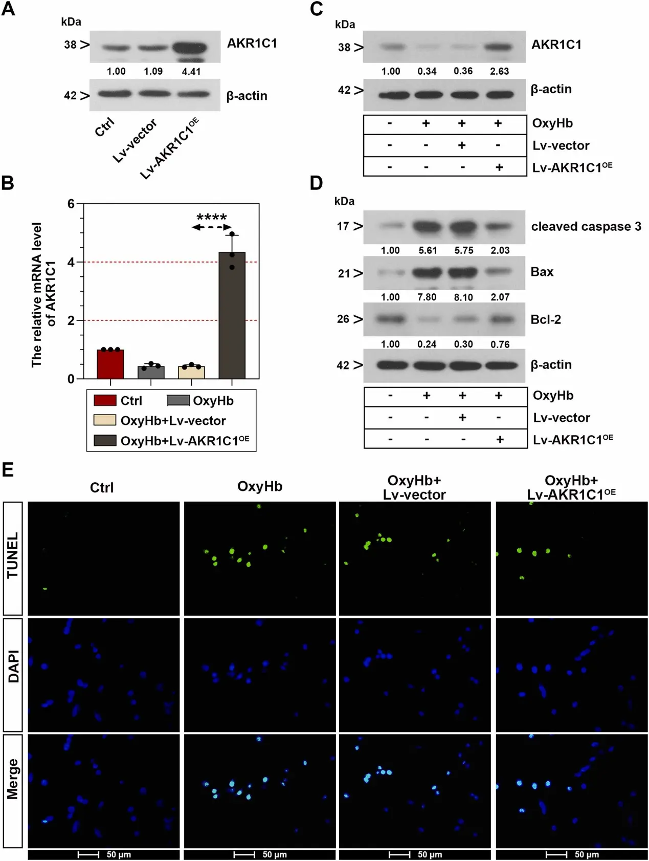
In this study, primary rat cortical neurons treated with oxygen hemoglobin (OxyHb) were applied to as an in vitro model. Following AKR1C1 overexpression, OxyHb-stimulated cells showed elevated AKR1C1 mRNA and protein levels (Fig. 1B-C). Consistent with the in vivo results, forced expression of AKR1C1 changed the protein amounts of the apoptosis-associated factors and reduced the number of TUNEL-labeled cells following OxyHb stimulation in neurons (Fig. 1D-E).
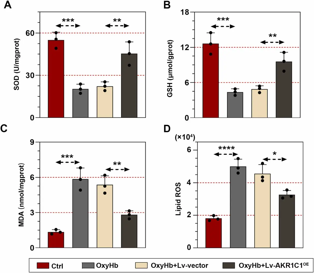
To verify whether AKR1C1 inhibited ferroptosis in neurons, we further measured the levels of oxidative stress biomarkers. In OxyHb-triggered neurons, the upregulation of AKR1C1 led to an increase in SOD activity and the level of GSH (Fig. 2A-B), as well as the decrease of MDA level and intracellular ROS level (Fig. 2C-D).
Ask a Question
Write your own review
- You May Also Need
Description: The thoracic aorta is located in the chest cavity and gives off arteries that branch to the esophagus, pericardium, lungs, and trachea. The thoracic aorta can be subdivided into the ascending aorta, ...
Description: Rat Podocytes are isolated from normal rat kidney. The cells are characterized by immunofluorescence with antibodies specific to podocin, Ang1, Nephrin, ACTN4, NPHS2. T25 flasks is required for cell ...
Description: Rat Bronchial Smooth Muscle Cells are isolated from normal rat bronchi tissue. Rat Bronchial Smooth Muscle Cells are characterized by immunofluorescence with antibodies specific to alpha-actin. T25 ...
Description: Rat Lung Epithelial Cells are isolated from normal rat lung tissue. The cells are characterized by immunofluorescence with antibodies specific to CK-18, CK-19. T25 flasks is required for cell ...
Description: Rat Vein Endothelial Cells from Creative Bioarray are isolated from inferior vena cava tissue of 6-8 week old laboratory Sprague-Dawley rat. Rat Vein Endothelial Cells are grown in T75 tissue culture ...

Description: Rat Corneal Epithelial Cells from Creative Bioarray are isolated from tissue of 1-day-old neonatal laboratory Sprague–Dawley Rats. Rat Corneal Epithelial Cells are grown in T25 tissue culture flask ...