
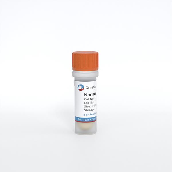
Normal Human Dermal Fibroblasts
Cat.No.: CSC-C4123X
Species: Human
Source: Dermis; Skin
Cell Type: Fibroblast
- Specification
- Background
- Scientific Data
- Q & A
- Customer Review
Cell Features:
HDFn are cryopreserved as primary cells. Cells are isolated from neonatal human foreskin and expanded in culture vessels once before cryopreservation.
HDFa are cryopreserved as primary cells. Cells are isolated from adult human skin and expanded in culture vessels once before cryopreservation.
Human Lung Fibroblasts are cryopreserved as secondary cells cells are isolated from human lungs and expanded in culture vessels twice before cryopreservation.
Fibroblasts can be grown in serum-free, animal protein-free, phenol red-free, and antimicrobial free conditions when cultured in FibroLife Serum-Free Medium.
Fibroblasts are extensively tested to meet quality standards and exhibit optimal performance.
Creative Bioarray's Fibroblasts are suitable for use as feeder layers as xeno-free or non-xeno free cultures for human embryonic stem cells and other cell culture applications requiring feeder layers.
Normal Human Dermal Fibroblasts (NHDF) are primary stromal cells isolated from normal human dermal tissue. They maintain the skin’s connective tissue environment by synthesizing and remodeling extracellular matrix (ECM), including collagen and fibronectin, and by producing signaling molecules that influence neighboring cells. These functions make NHDF a biologically relevant model for investigating dermal homeostasis, tissue repair, and matrix regulation.
NHDF provide a reference for examining changes associated with injury, fibrosis, and cellular aging. Their measurable responses—including proliferation, migration, matrix production, and contraction—support complementary assays that connect molecular signaling with functional outcomes. Under appropriate conditions, transforming growth factor-β can induce myofibroblast differentiation and increase fibrosis-associated gene expression, enabling controlled investigation of fibroblast activation and candidate antifibrotic interventions.
NHDF also offer considerable flexibility across experimental formats. They can be studied in conventional monolayers, incorporated into three-dimensional collagen matrices, or combined with keratinocytes to construct human skin equivalents. These systems enable investigation of epithelial–mesenchymal communication, matrix-dependent cellular behavior, and wound re-epithelialization. More complex models incorporating additional cell types further support studies of inflammatory and fibrotic interactions within a human tissue context.
Cytotoxicity Evaluation of Microbial Sophorolipids and Glucolipids Using Normal Human Dermal Fibroblasts (NHDF) In Vitro
Fibroblasts are a key cellular component for skin wound healing due to their capacity to secrete pro-regenerative growth factors, ECM proteins, and immunomodulators. Sophorolipids (SL), sophorosides (SS) and glucolipids (G), mono-unsaturated (C18:1) or saturated (C18:0), constitute a novel sustainable class of bio-based chemicals with interesting physicochemical characteristics, which allow them to form soft diverse structures from hydrogels to vesicles, micelles or complex coacervates with potential interest in skin regeneration applications.
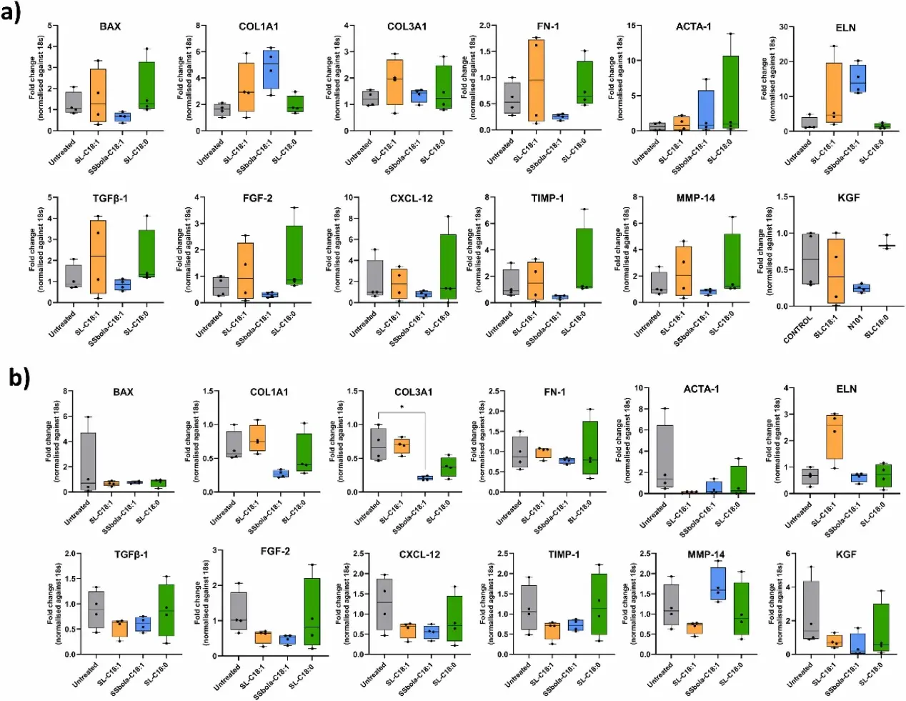
In this study, the cytocompatibility of a broad set of molecules from this family was tested on normal human dermal fibroblasts (NHDF). The results show that, up to an upper threshold (0.1 % w/v), the microbial glycolipids (SL-C18:1, G-C18:1, SSbola-C18:1, SL-C18:0 and G-C18:0) under study were able to sustain cell growth. Furthermore, the least cytotoxic glycolipids (SL-C18:1, SSbola-C18:1, SL-C18:0) were selected to study their potential to promote wound healing by measuring the gene expression of several key skin regeneration markers (i.e. collagen, elastin, transforming growth factor β, fibroblast growth factor …) using qPCR. Unfortunately, none of these glycolipids modulated the gene expression of molecules involved in tissue repair. However, this study aims to encourage the community to test this novel class of molecules for novel high-end biomedical applications.
Ask a Question
Write your own review
Description: RFP Expressing Human Dermal Lymphatic Microvascular Endothelial Cells (RFP-HDLMvECs) provided by Creative Bioarray are are puromycin-selected from Human Dermal Lymphatic Microvascular Endothelial ...
Description: Creative Bioarray's normal Human Dermal Fibroblasts-Neonatal, Xeno-Free (HDFn-XF), when grown in Creative Bioarray's LIFib Xeno-Free Complete Medium, provide an ideal xeno-free culture system to ...
Description: Neonatal Human Dermal Fibroblasts (NHDF) are selected by first isolating fibroblasts from the dermis of neonatal skin and then establishing a primary fibroblast culture. Upon completion of the ...
Description: Fibroblasts are mesenchymal cells derived from the embryonic mesoderm. They have been extensively used for a wide range of cellular and molecular studies. This is mainly because they are one of ...
Description: GFP and Luciferase Expressing Human Dermal Fibroblasts (Neonatal) (GFP-Luc-HNDFCs-Neo) provided by Creative Bioarray are puromycin-selected from Human Dermal Fibroblasts infected with lentivirus ...
Description: GFP Expressing Human Dermal Fibroblasts (neonatal) (GFP-HDFCs-Neo) provided by Creative Bioarray are puromycin-selected from Human Dermal Fibroblasts infected with lentivirus expressing GFP. ...