
Human Alveolar Epithelial Cells
Cat.No.: CSC-C9223J
Species: Human
Source: Lung
Cell Type: Epithelial Cell
- Specification
- Background
- Scientific Data
- Q & A
- Customer Review
The mature alveolar epithelium consists of type I and type II alveolar epithelial cells (AECI and AECII), also known as type I and type II pneumocytes, which account for about 96% and 4% respectively of the surface, although their numbers are roughly similar. AECIs, which are membranous flat cells usually found overlying the capillaries, provide the thin surface of the alveolus. The AECIIs are large cuboidal cells located in the corners of the alveoli, with a characteristic morphology of lamellar bodies and apical microvilli.
Functions of Alveolar Epithelial Cells
Gas exchange - The most notable feature of the alveolar region is its considerable surface of contact between air and blood, which is required for the huge gas exchange function adapted to the human body's oxygen demand.
Barrier function - One of the major roles of the lung epithelium is to serve as a physical barrier between the lumen and the underlying submucosa, protecting the tissue from bacteria, virus, allergens, and other noxious substances.
Immunomodulatory function - In addition to their function of mechanical barrier against pathogens and various inhaled environmental materials, AECs participate actively in the immune response of the lung. From the numerous studies focused on the inflammatory response of the alveolar epithelium and the identification of the myriad of molecules that may be generated under various insult conditions, distinct roles of AECIs and AECIIs begin to emerge.
Alterations in the function of AECs are a hallmark of a large variety of acute and chronic lung diseases, including lung cancer. The function of these cells can be studied in human lung tissue, in animal models and in cell culture. Cell culture has the advantage that isolated cells can be exposed to disease-relevant stimuli under controlled conditions, and can be manipulated by a variety of techniques, including genetic editing.
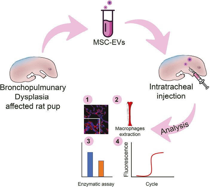
Extracellular Vesicles From Mesenchymal Umbilical Cord Cells Exert Protection Against Oxidative Stress and Fibrosis in a Rat Model of Bronchopulmonary Dysplasia
Background
This study investigates clinical-grade mesenchymal stromal cell-derived extracellular vesicles (MSC-EVs) from human umbilical cord in a hyperoxia-induced rat model of bronchopulmonary dysplasia. Intratracheal MSC-EVs reached lung and brain, reduced oxidative DNA damage and protein carbonylation, improved alveolar epithelial function, and attenuated collagen deposition and profibrotic signaling. In vitro, MSC-EVs suppressed TGFβ-induced macrophage-to-myofibroblast transition and protected alveolar epithelial cells from H2O2-induced DNA oxidation more effectively than vitamin C. These results suggest MSC-EVs as a potential therapy to prevent BPD progression.
Key Findings
Clinical-grade MSC-EVs were ~106 nm, expressed CD9/CD63/CD81, and after intratracheal delivery were detected in rat pup lungs and brain at 3 and 24 hours, with lung epithelial co-localization. In hyperoxia-induced BPD, MSC-EVs reduced oxidative stress markers 8-oxo-dG in alveolar ATI/ATII cells and brain astrocytes, and lowered protein carbonylation. They restored ATI/ATII balance, improved surfactant protein C and glycosaminoglycan expression, and ameliorated mitochondrial ultrastructure and Sod2 expression. Hyperoxia caused lung fibrosis with increased collagen deposition and Tgfβ1/αSMA; MSC-EVs significantly reduced collagen and blocked CD68+/αSMA+ macrophage accumulation, while increasing resident alveolar macrophages. In vitro, MSC-EVs suppressed TGFβ-induced macrophage-to-myofibroblast transition and collagen I production. In H2O2-damaged human alveolar epithelial cells, MSC-EVs reduced 8-oxo-dG more effectively than vitamin C, restoring proliferation. Thus, intratracheal MSC-EVs protect against oxidative stress, epithelial dysfunction, and fibrosis in experimental BPD.
Contribution
Creative Bioarray provided the human alveolar epithelial cell line CSC-C9223J and Supercult Alveolar Epithelial Cell Medium used to establish the H2O2-induced oxidative stress model. These reagents enabled assessment of MSC-EV antioxidant protection and DNA oxidation repair in lung epithelial cells, supporting the in vitro mechanistic validation.
DPP4 Inhibition Expands Human and Mouse AEC2s
To identify mechanisms for expanding AEC2s, we envisioned performing a high-content imaging screen, evaluating markers of proliferation in AEC2s in response to compound treatment. To overcome the technical difficulty of obtaining enough primary AEC2s to screen a large chemical library, we established a miniaturized screening assay using commercially available primary human small airway epithelial cells (SAECs). SAECs from preselected batches with high AEC2 signatures were used at early passage to best preserve high AEC2 character.
Consistent with the literature, the growth factors EGF, IGF-1, and FGF-10 promoted proliferation of this cell type. In addition, four structurally distinct FDA-approved gliptins- sitagliptin, saxagliptin, alogliptin, and linagliptin, as well as several clinical-stage DPP4 inhibitors induced a robust proliferative response in SAECs. Of these, the dipeptidyl peptidase 4 (DPP4) inhibitor, NVP-728, was the most active and displayed minimal cytotoxicity (Fig. 1).
 Fig. 2. DPP4 inhibitors selectively expand AEC2s (Shao, Sida, et al. 2024).
Customized Culture Medium for In Vitro Growth of Human Alveolar Epithelial Cells (AECs)
The researchers defined a small-molecule cocktail (SMC) medium, which was supplemented with Jagged-1 peptide (JAG-1), recombinant human Noggin protein (hNoggin), SB431542, recombinant human fibroblast growth factor (hFGF)-10 protein, hFGF-7, CHIR99021, and Y-27632, as an initial step in designing a standard method for long-term 2D culture of AECs. They confirmed the morphological changes to identify the expression of the epithelial cell adhesion molecule (EpCAM) and Vimentin-specific markers of epithelial and mesenchymal cells, respectively-to determine whether the SMC medium supports in vitro growth of AECs (Fig. 2a). They also evaluated the necessity of various supplements in the SMC medium by removing single additives, i.e., –JAG-1, –hNoggin, –SB431542, –hFGF-10, –hFGF-7, –CHIR99021, and –Y-27632, to evaluate the necessity of various supplements in the SMC medium. The results provide important insights into the support of SMC medium for the in vitro culture of the AECs, which emphasizes the realization of the stable culture of AECs (Fig. 2).
 Fig. 3. Development of customized human alveolar epithelial cell culture medium (Yang, Jia-Wei, et al. 2022).
Human Alveolar Epithelial Cells can be expanded for 3-7 passages at a split ratio of 1:2 or 1:3 under the cell culture conditions specified by Creative Bioarray. Repeated freezing and thawing of cells is not recommended.
No, the cells are isolated from healthy donor.
Ask a Question
Write your own review
Description: Human Pulmonary Artery Fibroblasts are isolated from normal human pulmonary artery.
Description: Primary Human Fetal Lung Fibroblast Cells were initiated from human lung tissue (normal 25 week gestation).These cells were originated using Complete Serum-Free Medium Kit With SuperFuel™, are ...
Description: GFP-Human Lung Airway Smooth Muscle Cells (GFP-HLAWSMCs) provided by Creative Bioarray are puromycin-selected from primary human lung airway smooth muscle cells infected with lentivirus expressing ...
Description: ACE2-GFP Expressing Human Pulmonary Artery Endothelial Cells (ACE2-GFP-HPAECs) provided by Creative Bioarray are puromycin-selected from Human Pulmonary Artery Endothelial Cells infected with ...
Description: The human type 1 alveolar epithelial cells from Creative Bioarray are isolated from human alveolar tissue. These cells are shipped at passage 2 and maintained in SuperCult® human type 1 alveolar ...
Description: Human Lung Tumor-Associated Endothelial Cells (Adenocarcinoma) from Creative Bioarray are isolated from human lung tumor tissue. Human Lung Tumor-Associated Endothelial Cells (Adenocarcinoma) are ...