
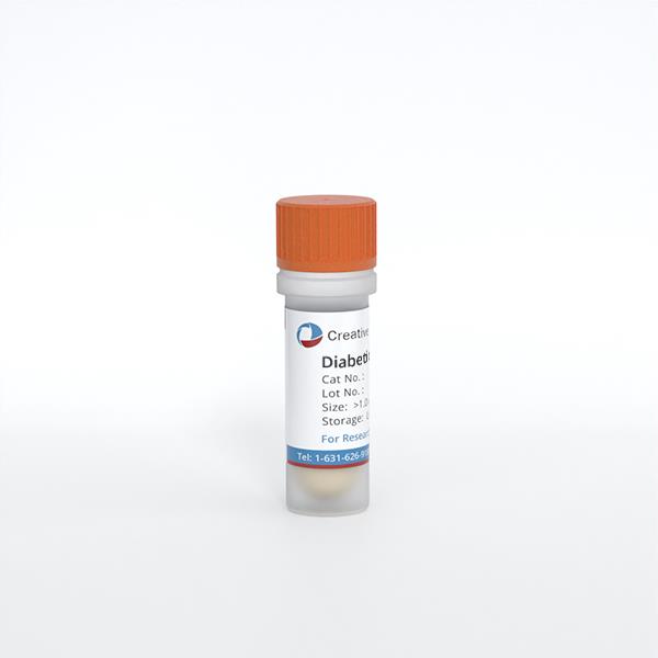
Diabetic Mouse Cardiac Microvascular Endothelial Cells
Cat.No.: CSC-C9300J
Species: Mouse
Source: Heart
Cell Type: Endothelial Cell; Microvascular Cell
- Specification
- Background
- Scientific Data
- Q & A
- Customer Review
Diabetic Mouse Cardiac Microvascular Endothelial Cells are primary endothelial cells isolated from the cardiac microvasculature of diabetic mice. They provide a disease-relevant model for investigating endothelial mechanisms underlying coronary microvascular dysfunction and diabetic cardiomyopathy. Their principal advantage is the combination of cardiac tissue origin and prior exposure to the diabetic environment in vivo, enabling investigation of disease-associated cellular alterations beyond responses to short-term glucose treatment alone.
These cells are particularly valuable for studying the relationship between metabolic stress and endothelial dysfunction. Research using cardiac microvascular endothelial cells isolated from db/db mice has demonstrated increased intracellular reactive oxygen species, reduced glycolytic reserve, and altered expression of genes involved in mitochondrial energy metabolism. Reduced proliferation and increased apoptosis were also observed, illustrating their utility for examining how diabetes affects endothelial survival and microvascular maintenance.
When compared with appropriately matched non-diabetic controls, these cells facilitate evaluation of disease-dependent responses and help connect cellular findings with cardiac vascular outcomes in vivo. Together, their tissue specificity and diabetic donor background make them a valuable resource for mechanistic and preclinical studies of diabetes-associated cardiac microvascular disease.
Adipsin Mitigates Cardiac Microvascular Injury in Diabetic Cardiomyopathy
This study was designed to investigate the potential role of Adipsin in the regulation of diabetic microvascular complications, as well as the underlying mechanism involved. Experimental diabetes was induced in mice with adipose tissue-specific Adipsin overexpression (AdipsinLSL/LSL-Cre) and their respective control (AdipsinLSL/LSL). In addition, cultured cardiac microvascular endothelial cells (CMECs) were subjected to high glucose/palmitic acid (HG + PA) treatment to simulate diabetes for a mechanistic approach. CMECs were treated with exosomes secreted by different mouse adipose tissues (AdipsinLSL/LSL and AdipsinLSL/LSL-Cre mice).
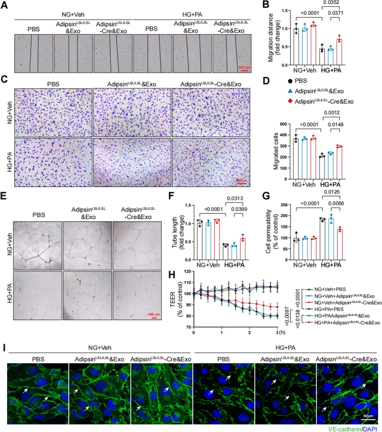
Adipsin-enriched exosomes were taken up by CMECs, inhibited CMECs apoptosis, and increased CMECs proliferation under HG + PA treatment. Adipsin-enriched exosomes also accelerated wound healing, rescued cell migration defects, and promoted tube formation in response to HG + PA challenge. Furthermore, Adipsin-enriched exosomes maintained adherens junctions at endothelial cell borders and reversed endothelial hyperpermeability disrupted by HG + PA insult. Together, these findings favor the vital role of Adipsin in the regulation of CMECs adherens junctions integrity, revealing its promises as a treatment target against diabetic coronary microvascular dysfunction.
Ask a Question
Write your own review
- You May Also Need


Description: C57BL/6-GFP Mouse Skeletal Muscle Microvascular Endothelial Cells from Creative Bioarray are isolated from C57BL/6-Tg (CAG-EGFP) 1Osb/J mouse skeletal muscle tissue of pathogen-free laboratory mice. ...
Description: eNOS KO Mouse Stomach Epithelial Cells from Creative Bioarray are isolated from stomach tissue of pathogen-free laboratory mice. eNOS KO Mouse Stomach Epithelial Cells are grown in a T25 tissue ...
Description: eNOS KO Mouse Liver Fibroblasts from Creative Bioarray are isolated from liver tissue of pathogen-free laboratory mice. eNOS KO Mouse Liver Fibroblasts are grown in T75 tissue culture flasks ...

Description: C57BL/6-GFP Mouse Corneal Epithelial Cells from Creative Bioarray are isolated from C57BL/6-GFP-Tg(CAG-EGFP)1Osb/J mouse corneal tissue of pathogen-free laboratory mice. C57BL/6-GFP Mouse Corneal ...

Description: BALB/c Mouse Retinal Microvascular Endothelial Cells from Creative Bioarray are isolated from retinal tissue of pathogen-free laboratory mice. BALB/c Mouse Retinal Microvascular Endothelial Cells are ...