Rat Oviduct Epithelial Cells
Cat.No.: CSC-C5046S
Species: Rat
Source: Oviduct
Cell Type: Epithelial Cell
- Specification
- Background
- Scientific Data
- Q & A
- Customer Review
Rat Oviduct Epithelial Cells (rOECs)from Creative Bioarray are isolated from the rat oviduct tissue. The method we use to isolate rOECs was developed based on a combination of established and our proprietary methods. The rOECs are characterized by immunofluorescence with antibodies specific to cytokeratin-18 (CK-18). Each vial contains 0.5x10^6 cells per ml and is delivered frozen.
Rat Oviduct Epithelial Cells (ROECs) constitute the functional lining of the maternal reproductive tract, serving as the biological interface for critical early reproductive events, including sperm capacitation, fertilization, and initial zygotic cleavage. Unlike immortalized lines, primary ROECs preserve the complex physiological responsiveness and polarized architecture required to simulate the oviductal microenvironment in vitro.
- Biphasic Cellular Functionality: ROECs maintain a sophisticated balance of ciliated cells, responsible for directed ovum and embryo transport, and secretory cells, which produce the oviduct-specific fluid rich in growth factors and proteins (e.g., OVGP1). This dual-population model is essential for studying the biochemical modulation of gamete maturation.
- Hormonal and Mechanical Fidelity: These cells exhibit high sensitivity to estrogen and progesterone fluctuations, accurately reflecting the cyclic changes of the estrous cycle. This makes ROECs a superior model for investigating endocrine-disrupting chemicals (EDCs) and the pharmacology of hormonal contraceptives.
- Optimal Environment for Pre-implantation Research: ROECs are a premier tool for co-culture systems aimed at overcoming the "in vitro developmental block". By providing essential paracrine signals and metabolic support, they significantly enhance embryo quality and developmental competence compared to synthetic media.
- High Translational Relevance in Rodent Models: The rat model offers a larger tissue yield and more distinct physiological phases than mouse models, facilitating more complex surgical simulations, proteomic analyses, and the study of pelvic inflammatory disease (PID) or tubal transport disorders.
Our Rat Oviduct Epithelial Cells are rigorously validated for purity using markers such as Cytokeratin 18 (CK18) and PAX8. By providing a standardized, "reproduction-ready" cellular system, this product minimizes experimental noise and accelerates discovery in reproductive toxicology, fertility research, and gynecological oncology. It is an indispensable asset for pharmaceutical R&D and academic centers focused on the frontiers of developmental biology.
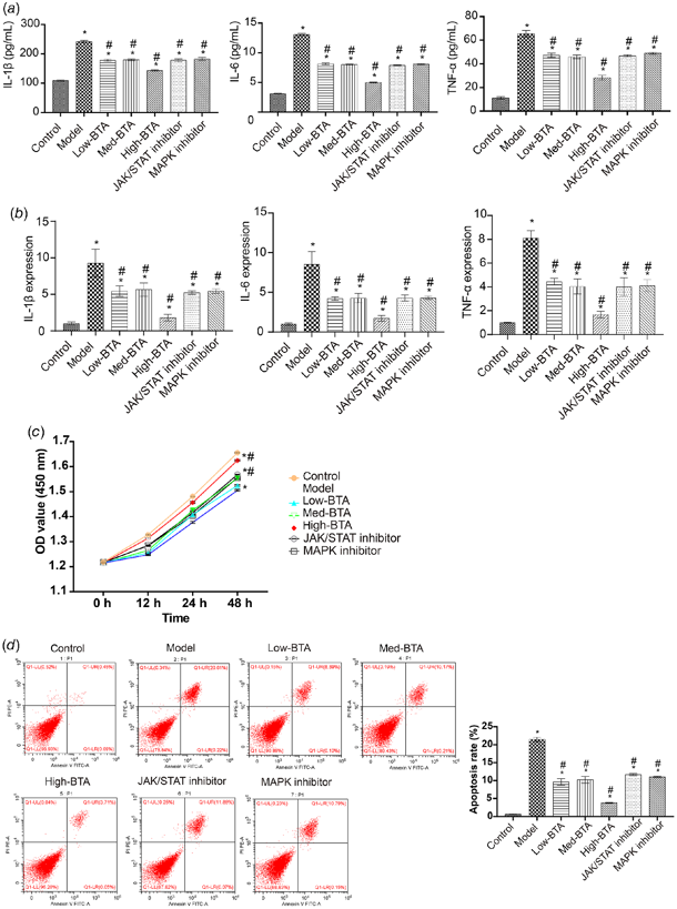
Betulonic Acid Regulates Oviduct Epithelial Cell Inflammation Through the TLR4, MAPK, and JAK/STAT Signaling Pathways
This study aimed to study the active effect and mechanism of betulonic acid (BTA) on tubal inflammatory infertility.
An inflammatory model was established in isolated rat oviduct epithelial cells. Immunofluorescence of cytokeratin 18 was performed in cells. The therapeutic effect of BTA on cells was observed. Subsequently, we added JAK/STAT inhibitor AG490 and MAPK inhibitor U0126 and measured the levels of inflammatory factors via enzyme-linked immunosorbent assay and qRT-PCR.
Betulonic acid inhibited the activation of TLR4 and NF-κB signaling pathways, and significantly downregulated IL-1β, IL-6, and TNF-α, with high doses being the most effective. Furthermore, high-dose BTA promoted the proliferation of oviduct epithelial cells and inhibited apoptosis. In addition, BTA inhibited the activation of JAK/STAT signaling pathway to perform effectively in oviduct epithelial cells inflammation. The addition of AG490 led to the inhibition of the JAK/STAT signaling pathway. BTA also inhibited the activation of MAPK signaling pathway in oviduct epithelial cells inflammation. Under U0126 treatment, the inhibition of proteins in MAPK pathway by BTA was weakened.
Ask a Question
Write your own review
- You May Also Need

Description: The thoracic aorta is located in the chest cavity and gives off arteries that branch to the esophagus, pericardium, lungs, and trachea. The thoracic aorta can be subdivided into the ascending aorta, ...

Description: Rat Podocytes are isolated from normal rat kidney. The cells are characterized by immunofluorescence with antibodies specific to podocin, Ang1, Nephrin, ACTN4, NPHS2. T25 flasks is required for cell ...

Description: Rat Bronchial Smooth Muscle Cells are isolated from normal rat bronchi tissue. Rat Bronchial Smooth Muscle Cells are characterized by immunofluorescence with antibodies specific to alpha-actin. T25 ...

Description: Guinea Pig Endothelial Cells from Creative Bioarray are isolated from guinea pig tissue. Prior to shipping, cells at passage 2 are detached from flasks and immediately cryopreserved in vials. Each ...

Description: Rat Lung Epithelial Cells are isolated from normal rat lung tissue. The cells are characterized by immunofluorescence with antibodies specific to CK-18, CK-19. T25 flasks is required for cell ...

Description: Rat Vein Endothelial Cells from Creative Bioarray are isolated from inferior vena cava tissue of 6-8 week old laboratory Sprague-Dawley rat. Rat Vein Endothelial Cells are grown in T75 tissue culture ...