
Rat Enteric Glial Cells
Cat.No.: CSC-C5103S
Species: Rat
Source: Intestine
Cell Type: Glial Cell
- Specification
- Background
- Scientific Data
- Q & A
- Customer Review
Rat Enteric Glial Cells from Creative Bioarray are isolated from the rat intestine tissue. The method we use to isolate Rat Enteric Glial Cells was developed based on a combination of established and our proprietary methods. The Rat Enteric Glial Cells are characterized by immunofluorescence with antibodies specific to glial fibrillary acidic protein(GFAP). Each vial contains 0.5x10^6 cells per ml and is delivered frozen.
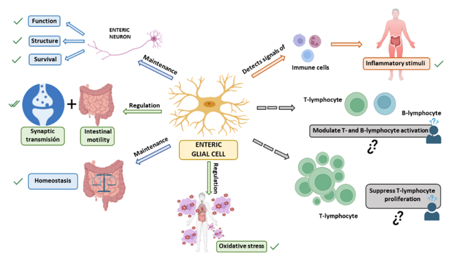
Rat Enteric Glial Cells are enteric glial cells isolated from the enteric nervous system of rats. The enteric nervous system (ENS) is a complex network of neurons found in the gastrointestinal tract, which modulates gut motility, secretion, blood flow, and gut-immune communication. Enteric glial cells are the most predominant non-neuronal cell type of the ENS and contribute to intestinal homeostasis and neuronal survival. Functionally and structurally similar to astrocytes of the central nervous system, enteric glial cells express glial fibrillary acidic protein (GFAP), S100β, and Sox10.
Primary cultured rat enteric glial cells serve as a tool to better understand gut neurobiology and gastrointestinal disorders. They can be used to model neuron-glia communication, intestinal inflammation, intestinal epithelial permeability, and gut-brain axis communication. Rat enteric glial cells are also often used to model gastrointestinal motility disorders, inflammatory bowel disease, and other forms of enteric neuropathy and intestinal damage. Rats are one of the most common species used to model gastrointestinal diseases, thus rat enteric glial cells serve as a useful experimental tool to study the workings of the ENS as well as study novel therapies aimed at modulating gut neuroimmune communication.
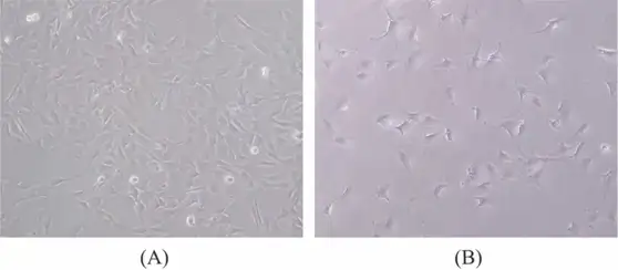
Microscopy-based Observation of EGCs Treated with Different Concentrations of Exogenous H2S
Hydrogen sulfide (H2S) is a gaseous signaling molecule influencing digestive and nervous system functions. Enteric glial cells (EGCs) regulate gastrointestinal motility within the enteric nervous system. Liu's team explored the dual effects of exogenous H2S on rat EGCs and its influence on apoptosis-related pathways and ion channels.
Control EGCs exhibited normal growth, flattened morphology, and extensive glial filament connections by 24 h. In contrast, 15 mM H2S treatment for 24 h inhibited growth, caused nuclear and cytoplasmic heterogeneity, retracted glial filaments, and disrupted intercellular junctions (Fig. 1). Low concentration (100 μM) showed no evident density changes. MTT assay was subsequently used for more accurate proliferation assessment.
Ask a Question
Write your own review

Description: The thoracic aorta is located in the chest cavity and gives off arteries that branch to the esophagus, pericardium, lungs, and trachea. The thoracic aorta can be subdivided into the ascending aorta, ...

Description: Rat Podocytes are isolated from normal rat kidney. The cells are characterized by immunofluorescence with antibodies specific to podocin, Ang1, Nephrin, ACTN4, NPHS2. T25 flasks is required for cell ...

Description: Rat Bronchial Smooth Muscle Cells are isolated from normal rat bronchi tissue. Rat Bronchial Smooth Muscle Cells are characterized by immunofluorescence with antibodies specific to alpha-actin. T25 ...

Description: Guinea Pig Endothelial Cells from Creative Bioarray are isolated from guinea pig tissue. Prior to shipping, cells at passage 2 are detached from flasks and immediately cryopreserved in vials. Each ...

Description: Rat Lung Epithelial Cells are isolated from normal rat lung tissue. The cells are characterized by immunofluorescence with antibodies specific to CK-18, CK-19. T25 flasks is required for cell ...

Description: Rat Vein Endothelial Cells from Creative Bioarray are isolated from inferior vena cava tissue of 6-8 week old laboratory Sprague-Dawley rat. Rat Vein Endothelial Cells are grown in T75 tissue culture ...