
Rat Endometrial Epithelial Cells
Cat.No.: CSC-C5048S
Species: Rat
Source: Endometrium; Uterus
Cell Type: Epithelial Cell
- Specification
- Background
- Scientific Data
- Q & A
- Customer Review
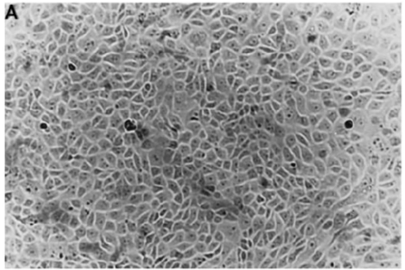
Rat Endometrial Epithelial Cells (rEECs) from Creative Bioarray are isolated from the rat uterine tissue. The method we use to isolate rEECs was developed based on a combination of established and our proprietary methods. The rEECs are characterized by immunofluorescence with antibodies specific to pan-cytokeratin (PCK). Each vial contains 0.5x10^6 cells per ml and is delivered frozen.
Rat Endometrial Epithelial Cells are primary cells isolated from the epithelial layer of the rat uterus. The endometrium is the inner lining of the uterus that undergoes cyclical changes in structure and function during the reproductive cycle. Endometrial epithelial cells line the luminal and glandular surfaces of the endometrium and are essential for creating a receptive uterine environment for embryo implantation, supporting early pregnancy, and maintaining uterine homeostasis. In vitro, Rat Endometrial Epithelial Cells exhibit epithelial cell morphology, including a polarized cobblestone appearance and the ability to form tight junctions that contribute to barrier function. These cells express epithelial markers and hormone-responsive receptors, such as estrogen and progesterone receptors, reflecting their responsiveness to cyclic hormonal regulation. They can modulate embryo-uterine communication and influence the local immune environment through regulated secretion of cytokines, growth factors, and extracellular matrix components.
Research utilizes Rat Endometrial Epithelial Cells to explore multiple dimensions of endometrial physiology and pathology which include hormonal signaling, uterine receptivity, and epithelial remodeling. These cells provide a model for investigating the mechanisms underlying implantation failure, endometrial inflammation, fibrosis, and reproductive toxicity. They are also employed in toxicological and pharmacological studies to assess the impact of drugs, endocrine-disrupting chemicals, and environmental factors on uterine epithelial function.
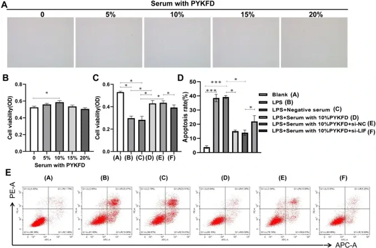
Effect of PYKFD on the Biological Behavior of REECs In Vitro
Tubal inflammation, endometritis, and uterine adhesions due to post-pelvic inflammatory disease (SPID) are significant causes of infertility. Chronic endometritis (CE) is a critical issue affecting women's reproductive health. This study investigates the effects of Pen Yan Kang Fu Decoction (PYKFD) on endometrial receptivity and repair via the LIF/JAK2/STAT3 signaling pathway in chronic endometritis infertility.
Serum from rats treated with high-dose PYKFD was prepared into media at 5%, 10%, 15%, and 20% concentrations for CCK-8, western blot, and flow cytometry apoptosis assays. Figure 1A and B show that 10% serum medium had the strongest effect on rat endometrial epithelial cells (REECs) viability. To verify LIF's role, si-LIF was used to block PYKFD induction. Figure 1C indicates that while 10% PYKFD serum promoted cell proliferation, si-LIF blocked this effect, whereas si-NC had no impact. Figures 4D and E show that LPS induced the most apoptosis in serum without PYKFD. After LIF silencing, the apoptosis reversal effect of PYKFD was reduced compared to the LPS + 10% PYKFD serum group. These results suggest that PYKFD's effects on endometrial epithelial cells involve LIF.
Ask a Question
Write your own review
- You May Also Need

Description: The thoracic aorta is located in the chest cavity and gives off arteries that branch to the esophagus, pericardium, lungs, and trachea. The thoracic aorta can be subdivided into the ascending aorta, ...

Description: Rat Podocytes are isolated from normal rat kidney. The cells are characterized by immunofluorescence with antibodies specific to podocin, Ang1, Nephrin, ACTN4, NPHS2. T25 flasks is required for cell ...

Description: Rat Bronchial Smooth Muscle Cells are isolated from normal rat bronchi tissue. Rat Bronchial Smooth Muscle Cells are characterized by immunofluorescence with antibodies specific to alpha-actin. T25 ...

Description: Guinea Pig Endothelial Cells from Creative Bioarray are isolated from guinea pig tissue. Prior to shipping, cells at passage 2 are detached from flasks and immediately cryopreserved in vials. Each ...

Description: Rat Lung Epithelial Cells are isolated from normal rat lung tissue. The cells are characterized by immunofluorescence with antibodies specific to CK-18, CK-19. T25 flasks is required for cell ...

Description: Rat Vein Endothelial Cells from Creative Bioarray are isolated from inferior vena cava tissue of 6-8 week old laboratory Sprague-Dawley rat. Rat Vein Endothelial Cells are grown in T75 tissue culture ...