
Rat Corpus Cavernosum Smooth Muscle Cells
Cat.No.: CSC-C5043S
Species: Rat
Source: Penis
Cell Type: Smooth Muscle Cell
- Specification
- Background
- Scientific Data
- Q & A
- Customer Review
Rat Corpus Cavernosum Smooth Muscle Cells (rCCSMCs) from Creative Bioarray are isolated from the rat penile tissue. The method we use to isolate rCCSMCs was developed based on a combination of established and our proprietary methods. The rCCSMCs are characterized by immunofluorescence with antibodies specific to α-SMA. Each vial contains 0.5x10^6 cells per ml and is delivered frozen.
Primary rat corpus cavernosum smooth muscle cells (CCSMCs), isolated by enzymatic dispersion from rat penile crura, constitute the principal cellular regulator of penile hemodynamics, governing both erection and detumescence. In culture, they faithfully retain their native phenotype, providing a powerful and physiologically authentic in vitro system for erectile biology research.
A principal advantage of rat CCSMCs is their robust preservation of a differentiated, contractile phenotype. The cells exhibit the classic spindle-shaped morphology and strongly express a full panel of smooth muscle markers, including α-smooth muscle actin, calponin, smooth muscle myosin heavy chain, and desmin. Functionally, they mount potent, dose-dependent contractions to α1-adrenergic agonists (e.g., phenylephrine) and endothelin-1, and relax in response to nitric oxide donors, natriuretic peptides, or direct activators of soluble guanylyl cyclase, enabling direct mechanistic dissection of the NO-cGMP and RhoA/ROCK signaling pathways central to penile erection. Critically, rat CCSMCs maintain high endogenous expression of phosphodiesterase type 5 (PDE5), the molecular target of sildenafil, making them an ideal platform for evaluating PDE5 inhibitors and novel relaxant agents.
The rat origin confers high availability, low cost, and seamless integration with established rodent models of erectile dysfunction—including diabetes, cavernous nerve injury, castration, and hypertension. This allows precise in vitro–in vivo correlation of molecular alterations such as phenotypic switching to a synthetic or fibrotic state, apoptosis, and oxidative stress. Collectively, primary rat CCSMCs offer an indispensable, translationally relevant platform for dissecting erectile physiology, elucidating the pathogenesis of erectile dysfunction, and screening candidate therapeutics.
Angiotensin (1-7) Exerts a Protective Effect on Rat Corpus Cavernosum Smooth Muscle Cells under High Glucose Conditions
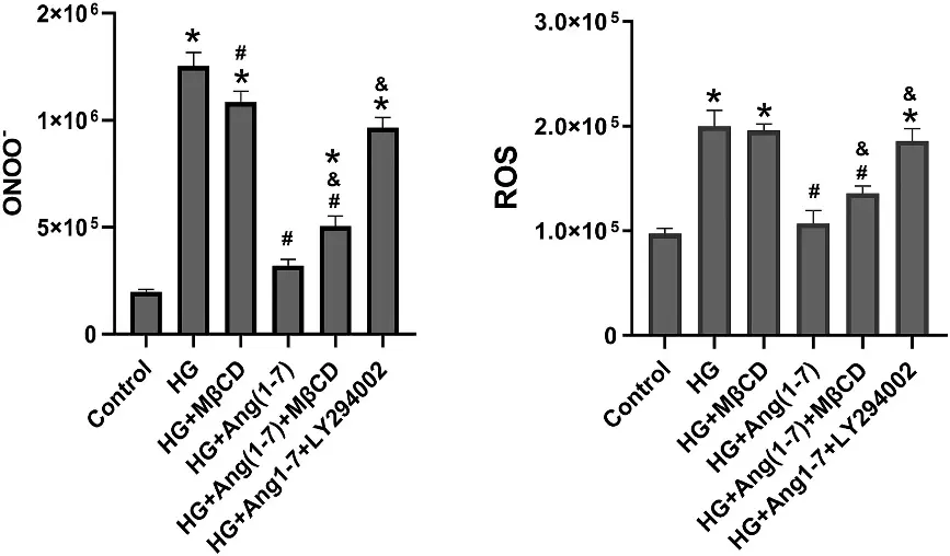
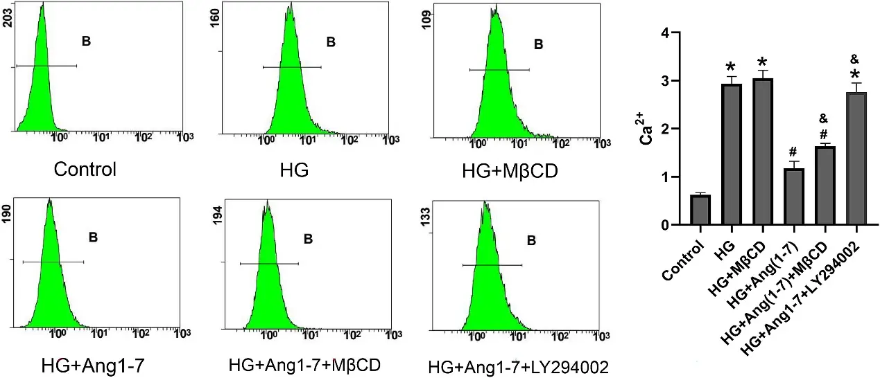
This study explored the role of Angiotensin (1-7) (Ang 1-7) and the regulatory mechanism underlying fibrosis in the corpora cavernosa. Primary rat corpus cavernosum smooth muscle cells (CCSMCs) were cultured under normal and high glucose (HG) with or without Ang 1-7. The protein levels of TGF-β1, Collagen I, TGF-β receptor-I (TβRI), and caveolin-1 (Cav-1) were evaluated by western blotting. Reactive oxygen species (ROS) and peroxynitrite (ONOO-) levels in cell culture supernatants were measured by enzyme-linked immunosorbent assay. Intracellular calcium content was determined by flow cytometry.
High glucose significantly increased the protein levels of TGF-β1 and Collagen I, triggered oxidative stress, modulated the AKT signaling pathway, and elevated intracellular calcium ion levels in CCSMCs. Angiotensin (1-7) treatment significantly attenuated HG-induced adverse effects in CCSMCs. The protective effects of Ang 1-7 against fibrosis in HG-exposed CCSMCs were associated with the downregulation of TGF-β1 levels, accompanied by the attenuation of oxidative stress. The Ang1-7-induced effects in CCSMC cells are mediated through the inhibition of the TGF-β1/AKT signaling pathway.
Ask a Question
Write your own review
- You May Also Need
Description: The thoracic aorta is located in the chest cavity and gives off arteries that branch to the esophagus, pericardium, lungs, and trachea. The thoracic aorta can be subdivided into the ascending aorta, ...
Description: Rat Podocytes are isolated from normal rat kidney. The cells are characterized by immunofluorescence with antibodies specific to podocin, Ang1, Nephrin, ACTN4, NPHS2. T25 flasks is required for cell ...
Description: Rat Bronchial Smooth Muscle Cells are isolated from normal rat bronchi tissue. Rat Bronchial Smooth Muscle Cells are characterized by immunofluorescence with antibodies specific to alpha-actin. T25 ...
Description: Guinea Pig Endothelial Cells from Creative Bioarray are isolated from guinea pig tissue. Prior to shipping, cells at passage 2 are detached from flasks and immediately cryopreserved in vials. Each ...
Description: Rat Lung Epithelial Cells are isolated from normal rat lung tissue. The cells are characterized by immunofluorescence with antibodies specific to CK-18, CK-19. T25 flasks is required for cell ...
Description: Rat Vein Endothelial Cells from Creative Bioarray are isolated from inferior vena cava tissue of 6-8 week old laboratory Sprague-Dawley rat. Rat Vein Endothelial Cells are grown in T75 tissue culture ...