
Canine Bone Marrow Macrophages
Cat.No.: CSC-C4841L
Species: Dog
Source: Bone Marrow
Cell Type: Macrophage
- Specification
- Background
- Scientific Data
- Q & A
- Customer Review
Never can cryopreserved cells be kept at -20 °C.
Canine Bone Marrow Macrophages are primary immune cells that are differentiated from bone marrow-derived monocytes isolated from dogs. These cells represent a physiologically relevant in vitro model of innate immune responses amenable to veterinary and translational research applications. They are mononuclear phagocytes that function as professional antigen-presenting cells and are critical for pathogen recognition and clearance, phagocytosis, antigen presentation, and inflammation.
In culture, Canine Bone Marrow Macrophages exhibit typical macrophage morphology, with adherent, spread-out cells displaying abundant cytoplasm and pseudopodia. Phenotypically, they express cell surface markers consistent with macrophages including CD14, CD11b, and MHC class II and can respond to numerous immunological stimuli (e.g., lipopolysaccharide [LPS], cytokines, microbial molecules). Cells can be stimulated with polarizing cytokines to induce M1- or M2-like phenotypes.
These cells have been used to study various infectious diseases, inflammation, cancer, and immunotoxicology. Canine Bone Marrow Macrophages can also be used as a model system for drug development of immunomodulatory therapeutics, vaccines, and canine-specific host-pathogen interactions. Many studies take advantage of these cells due to their primary nature and physiological relevance to the in vivo system.
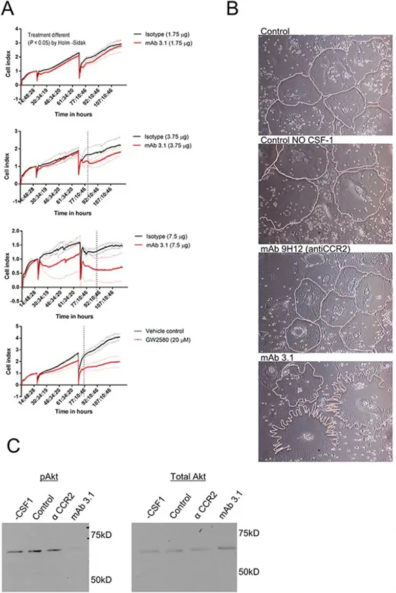
mAb 3.1 had an Inhibitory Effect on Canine Bone Marrow Macrophages Cultures
CSF-1R is a receptor mostly associated with the mononuclear phagocytic system. However, its expression within tumors has been linked with poor prognosis in both humans and dogs. CB Beirão et al. aimed to generate a canine-specific CSF-1R-blocking monoclonal antibody as the first step toward a veterinary anti-cancer biologic.
A murine hybridoma antibody (mAb3.1) was created that targeted the CSF-1R dimerization domain. mAb3.1 bound to fixed macrophages and inhibited proliferation of mononuclear phagocytic cells, but had weak reactivity against native glycoforms and off-target binding. In vitro, mAb 3.1 showed a significant inhibition of canine macrophage proliferation at a concentration of 1.25 μg/well in a real time proliferation assay (Holm-Sidak post hoc test). Trends of inhibition were seen at concentrations of 3.75 and 7.5 μg/well (Fisher post hoc test). Small molecule CSF-1R inhibitor GW2580 (20 μM) was used as a positive control and decreased macrophage survival compared to DMSO vehicle (Fig. 1A).
Ask a Question
Write your own review
- You May Also Need
Description: Dog Liver Endothelial Cells from Creative Bioarray are isolated from tissue of dog liver. Dog Liver Endothelial Cells are grown in T25 tissue culture flasks pre-coated with gelatin-based coating ...
Description: Canine Astrocytes from Creative Bioarray are isolated from canine brain tissue. The method we use to isolate canine astrocytes were developed based on a combination of established and our proprietary ...
Description: Canine Mammary Microvascular Endothelial Cells from Creative Bioarray are isolated from breast of pathogen-free laboratory Canine. Canine Mammary Microvascular Endothelial Cells are grown in T25 ...
Description: Canine Chondrocytes (CnC) provided by Creative Bioarray are isolated from normal canine articular cartilage tissue. The cells are frozen at passage 1 and each vial contains at least 0.5*10^6 cells. ...
Description: Canine Pancreatic Microvascular Endothelial Cells from Creative Bioarray are isolated from Pancreatic Microvascular of pathogen-free laboratory Canine. Canine Pancreatic Microvascular Endothelial ...
Description: Canine Prostate Microvascular Endothelial Cells from Creative Bioarray are isolated from prostate of pathogen-free laboratory Canine. Canine Prostate Microvascular Endothelial Cells are grown in T25 ...