
Human Skin Fibroblast-DMD
Cat.No.: CSC-C8158L
Species: Human
Source: Skin
Cell Type: Fibroblast
- Specification
- Scientific Data
- Q & A
- Customer Review
Desease: Duchenne Muscular Dystrophy (DMD)
Application: These cells can be used to study skin diseases such as wound healing, dermatitis, and other diseases that are expressed in fibroblast. In addition, these cells enable researchers to study the development of skin and production of inducible pluripotent stem cells.
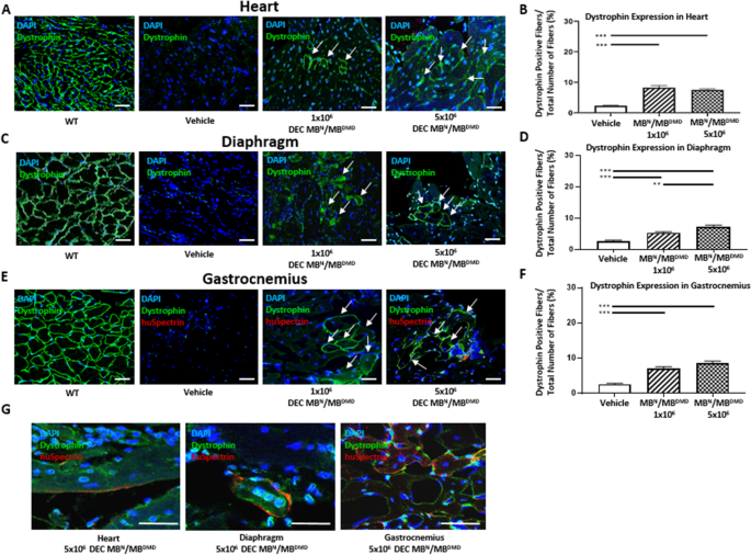
Long-Term Protective Effect of Human Dystrophin Expressing Chimeric (DEC) Cell Therapy on Amelioration of Function of Cardiac, Respiratory and Skeletal Muscles in Duchenne Muscular Dystrophy
Background
This study evaluates human dystrophin-expressing chimeric (DEC) cell therapy for Duchenne muscular dystrophy. DEC cells were created by fusing normal and DMD-affected donor myoblasts and administered systemically-intraosseously to mdx/scid mice. At 180 days, DEC therapy restored dystrophin expression and improved cardiac, respiratory, and skeletal muscle function. It reduced muscle inflammation, fibrosis, centrally nucleated fibers, and normalized fiber diameters. Functional gains included preserved ejection fraction/fractional shortening, better respiratory parameters, and increased grip strength. The findings support human DEC as a potential advanced therapy medicinal product for DMD.
Key Findings
Systemic-intraosseous human DEC therapy produced long-term engraftment and significantly increased dystrophin expression in heart, diaphragm, and gastrocnemius muscle at 180 days. Effects were dose-dependent, with 5×10^6 DEC cells often superior to 1×10^6. DEC therapy reduced inflammatory foci and fibrosis in all target organs, decreased centrally nucleated fibers in diaphragm and gastrocnemius, and shifted muscle fiber diameters toward normal. Functionally, DEC-treated mdx/scid mice showed maintained or improved ejection fraction, fractional shortening, stroke volume, and cardiac output; reduced enhanced pause and expiration time; and greater maximum stretch-induced contraction and grip strength. These benefits occurred without immunosuppression and after a single dose, supporting human DEC as a novel ATMP candidate capable of slowing or halting DMD progression.
Contribution
Creative Bioarray provided DMD-affected human myoblasts used as parent cells to generate human dystrophin-expressing chimeric (DEC) cells. These cells were fused with normal myoblasts, enabling the DEC therapy tested in mdx/scid mice and supporting the study’s preclinical assessment of long-term efficacy in DMD.
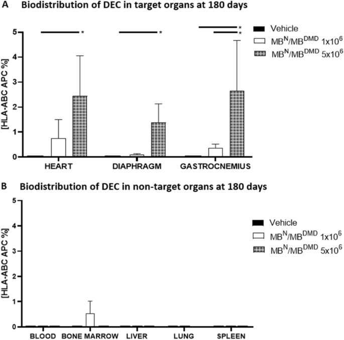
Long-Term Biodistribution and Safety of Human Dystrophin Expressing Chimeric Cell Therapy After Systemic-Intraosseous Administration to Duchenne Muscular Dystrophy Model
Background
This preclinical study assessed the long-term safety and biodistribution of human dystrophin-expressing chimeric (DEC) cell therapy in mdx/scid mice, a Duchenne muscular dystrophy model. After systemic-intraosseous administration of 1×10^6 or 5×10^6 DEC cells, assessments at 180 days included COMET assay, MRI, body/muscle/organ weights, flow cytometry for HLA-ABC, and immunofluorescence for dystrophin/human spectrin co-localization. DEC therapy caused no genotoxicity, tumorigenicity, or dose-related toxicity, showed dose-dependent engraftment in target muscles, and had negligible presence in non-target organs, supporting further clinical development.
Key Findings
At 180 days after systemic-intraosseous administration of human DEC cells to mdx/scid mice, COMET assay confirmed no DNA damage in parent or fused cells. No local complications, dose-related toxicity, or anaphylactic/immune reactions were observed after 1×10^6 or 5×10^6 DEC doses. MRI and autopsy revealed no tumor formation; body, muscle, and organ weights remained stable and comparable to vehicle controls, except one spontaneous murine sarcoma in an mdx/scid mouse. Flow cytometry showed dose-dependent HLA-ABC+ human DEC cells in target organs—heart, diaphragm, and gastrocnemius—with negligible presence in blood, bone marrow, lung, liver, and spleen. Immunofluorescence confirmed co-localization of dystrophin with human spectrin in target muscles, verifying human-origin dystrophin at 180 days. These results demonstrate long-term local and systemic safety, selective engraftment, and support DEC as an advanced therapy medicinal product for DMD.
Contribution
Creative Bioarray supplied DMD-affected human myoblasts, which served as parent cells for ex vivo fusion with normal human myoblasts to generate dystrophin-expressing chimeric (DEC) cells. This disease-relevant cell source was essential for creating and testing DEC therapy in the mdx/scid DMD model; no other contribution is reported.
Ask a Question
Write your own review
- You May Also Need
Description: Adult normal Primary Sebocytes are a vital research tool for a variety of applications from acne, seborrhoea and other sebaceous gland-related diseases studies in skin biology/cosmetic programs. ...
Description: Origin: HumanDesease: Psoriasis (PS)Application: These cells can be used to study skin diseases such as wound healing, dermatitis, and other diseases that are expressed in fibroblast. In addition, ...
Description: Origin: HumanDesease: AstrocytomaApplication: These cells can be used to study skin diseases such as wound healing, dermatitis, and other diseases that are expressed in fibroblast. In addition, these ...
Description: Origin: HumanDesease: Arteriovenous Malformation (AVM)Application: These cells can be used to study skin diseases such as wound healing, dermatitis, and other diseases that are expressed in ...
Description: Origin: HumanDesease: Rheumatoid Arthritis (RA)Application: These cells can be used to study skin diseases such as wound healing, dermatitis, and other diseases that are expressed in fibroblast. In ...

Description: Origin: HumanDesease: Diabetes Type IApplication: These cells can be used to study skin diseases such as wound healing, dermatitis, and other diseases that are expressed in fibroblast. In addition, ...