Rat Intestinal Mesenteric Vascular Endothelial Cells
Cat.No.: CSC-C4185X
Species: Rat
Source: Mesentery
Cell Type: Endothelial Cell
- Specification
- Background
- Scientific Data
- Q & A
- Customer Review
Rat Intestinal Mesenteric Vascular Endothelial Cells are endothelial cells harvested from the mesenteric microvasculature of the rat intestine. Endothelial cells line the interior surface of blood vessels in the mesenterium. They play a pivotal role in regulating vascular homeostasis and blood flow and mediate the trafficking of nutrients, metabolites and immune mediators to and from blood vessels and intestinal tissues. As part of the intestinal microcirculatory system, mesenteric vascular endothelial cells are an integral component of the gut-vascular barrier. The gut-vascular barrier along with the intestinal epithelial barrier serves to prevent pathogenic microbes and toxins from entering host circulation.
Microscopically, endothelial cells possess a cobblestone morphology when maintained in culture. In addition to displaying endothelial markers including CD31 (PECAM-1), von Willebrand factor (vWF) and vascular endothelial cadherin (VE-cadherin), endothelial cells take up acetylated low-density lipoprotein (Ac-LDL) and will form capillary like structures of tubules when cultured on Matrigel. Endothelial cells also exhibit high angiogenic potential.
Mesenteric Vascular endothelial cells function to regulate vascular permeability, leukocyte recruitment and trafficking, inflammation and angiogenesis. They have also been shown to play a role in intestinal immune homeostasis as well as intestinal perfusion. Perturbations in mesenteric endothelial cell function have been linked to several disease states including inflammatory bowel disease, ischemia-reperfusion injury, metabolic disease and portal hypertension.
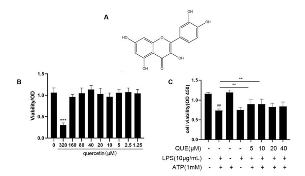
The Effect of Quercetin on the Survival Rate of RIMVECs
Intestinal mucosal microvascular endothelial cells (MECs) are involved in the pathogenesis of inflammatory bowel disease (IBD). Quercetin has been reported to have antitumor effects against gastrointestinal tract cancers, but its role in bacterial enteritis and diseases associated with pyroptosis remains unclear. Zhang et al. investigated the influence of quercetin on inflammatory injury and pyroptosis in rat intestinal mucosal vascular endothelial cells (RIMVECs).
Quercetin showed no effect on RIMVEC survival at 3-12 h treatment. After 24 h exposure to quercetin, only the 320 µM group displayed significantly decreased cell survival (P < 0.01). Groups treated with ≤80 µM quercetin did not cause RIMVEC toxicity (P < 0.01; Fig. 1A, B). Based on the CCK-8 assay findings and previous reports, the concentrations of quercetin used in subsequent experiments ranged from 5-40 µM.
Ask a Question
Write your own review

Description: The thoracic aorta is located in the chest cavity and gives off arteries that branch to the esophagus, pericardium, lungs, and trachea. The thoracic aorta can be subdivided into the ascending aorta, ...

Description: Rat Podocytes are isolated from normal rat kidney. The cells are characterized by immunofluorescence with antibodies specific to podocin, Ang1, Nephrin, ACTN4, NPHS2. T25 flasks is required for cell ...

Description: Rat Bronchial Smooth Muscle Cells are isolated from normal rat bronchi tissue. Rat Bronchial Smooth Muscle Cells are characterized by immunofluorescence with antibodies specific to alpha-actin. T25 ...

Description: Guinea Pig Endothelial Cells from Creative Bioarray are isolated from guinea pig tissue. Prior to shipping, cells at passage 2 are detached from flasks and immediately cryopreserved in vials. Each ...

Description: Rat Lung Epithelial Cells are isolated from normal rat lung tissue. The cells are characterized by immunofluorescence with antibodies specific to CK-18, CK-19. T25 flasks is required for cell ...

Description: Rat Vein Endothelial Cells from Creative Bioarray are isolated from inferior vena cava tissue of 6-8 week old laboratory Sprague-Dawley rat. Rat Vein Endothelial Cells are grown in T75 tissue culture ...