
Rabbit Coronary Artery Endothelial Cells
Cat.No.: CSC-C8664W
Species: Rabbit
Source: Artery
Cell Type: Endothelial Cell
- Specification
- Scientific Data
- Q & A
- Customer Review
High Magnesium and Sirolimus on Rabbit Vascular Cells—An In Vitro Proof of Concept
Background
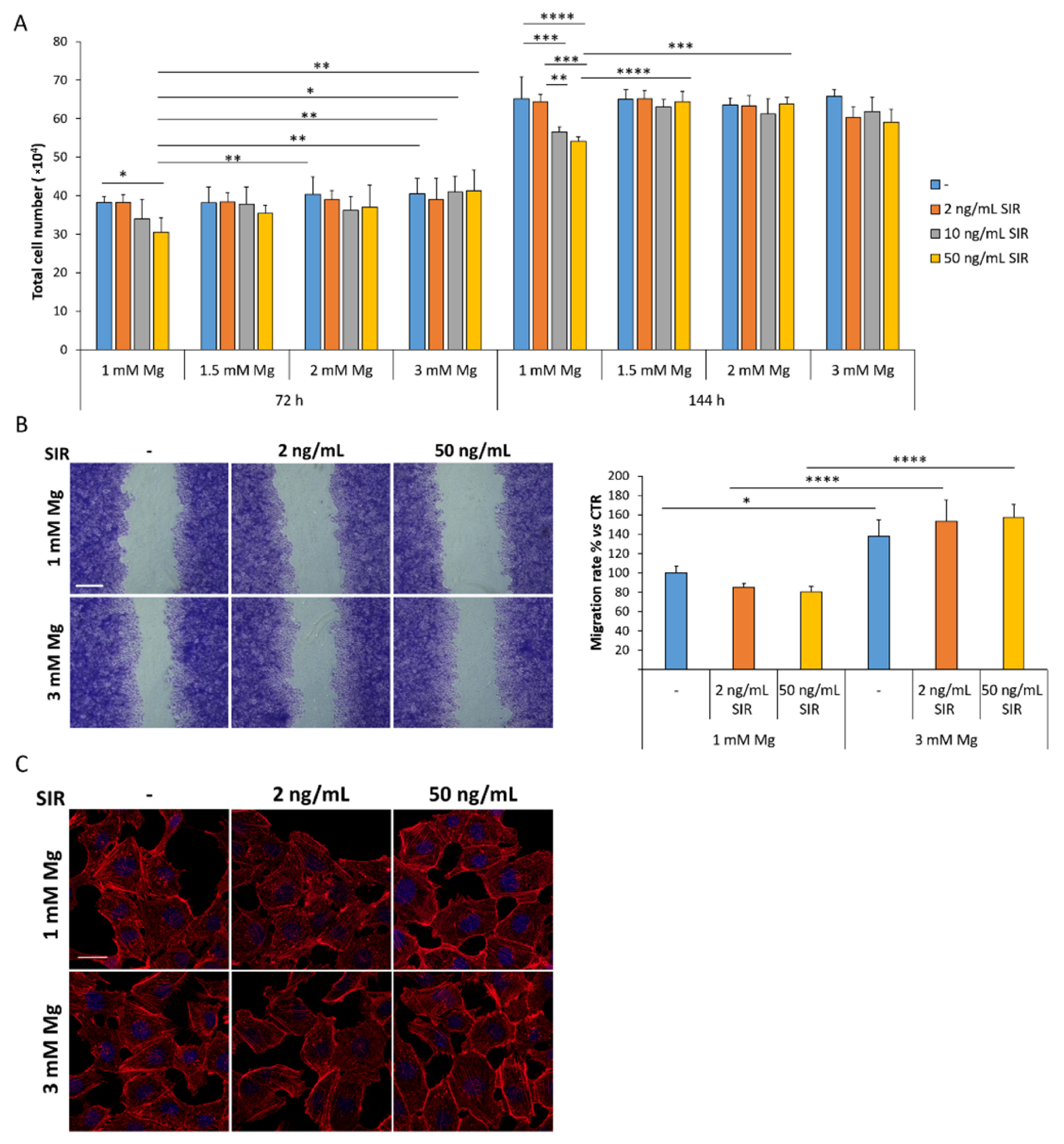
Drug-eluting bioresorbable magnesium scaffolds locally release magnesium, potentially interacting with sirolimus. Using rabbit coronary artery endothelial (rCAEC) and smooth muscle cells (rSMC), this study tested sirolimus with 1–3 mM extracellular magnesium. High magnesium prevented sirolimus-induced inhibition of rCAEC proliferation and stimulated migration, while it did not rescue sirolimus-mediated rSMC growth arrest and further reduced migration. Sirolimus and magnesium did not impair rSMC nitric oxide response. The results suggest local magnesium increases preserve endothelial function without compromising sirolimus's anti-restenotic effects on smooth muscle cells.
Key Findings
In rabbit coronary artery endothelial cells (rCAEC), sirolimus (2–50 ng/mL) inhibited proliferation only at physiological 1 mM magnesium; higher magnesium (1.5–3 mM) prevented this inhibition. At 3 mM magnesium, with or without sirolimus, rCAEC migration increased. Magnesium and sirolimus did not affect P-selectin; VCAM was downregulated at 72 h by 3 mM magnesium but upregulated at 144 h with 50 ng/mL sirolimus. Tissue factor decreased at 144 h with 3 mM magnesium plus sirolimus, while eNOS remained unchanged. In rabbit smooth muscle cells (rSMC), sirolimus strongly inhibited growth at all tested magnesium concentrations, and high magnesium (3 mM) did not rescue growth arrest; instead, it enhanced sirolimus's inhibition of migration. Neither sirolimus nor magnesium impaired the rSMC cGMP response to nitric oxide donor SNP. Overall, high local magnesium may favor endothelial recovery while maintaining sirolimus's anti-proliferative and anti-migratory effects on smooth muscle cells, supporting the biocompatibility of sirolimus-eluting magnesium scaffolds.
Contribution
Creative Bioarray provided primary rabbit coronary artery endothelial cells (rCAEC) and smooth muscle cells (rSMC) used in all experiments. These cells were expanded for three population doublings following the manufacturer's instructions, enabling assessment of sirolimus and magnesium effects in vitro.
Ask a Question
Write your own review

Description: Rabbit Hepatocytes are derived from the liver of New Zealand White Rabbit.
Description: Rabbit Pulmonary Artery Smooth Muscle Cells are isolated from pulmonary artery of New Zealand White Rabbit.
Description: Enteric nervous system glial cells resemble central nervous system astrocytes. The enteric nervous system is widely distributed throughout the intestinal tract in a network structure. Enteric glia ...
Description: The aortic arch is the top part of the main artery carrying blood away from the heart. It is the connection between the ascending and descending aorta, and its central part is formed by the left 4th ...
Description: The synovium secretes synovial fluid, which plays an important role in joint movement. The normal synovium has two layers, a thin cellular layer (luminal layer) and a vascular layer (subintima). ...
Description: Rabbit Prostate Fibroblasts are isolated from prostate tissue of New Zealand White Rabbit.