
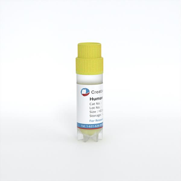
Human Skin Cells (Dermal Fibroblasts) (GM)
Cat.No.: CSC-C3570
Species: Human
Source: Dermis; Skin
Cell Type: Fibroblast
- Specification
- Background
- Scientific Data
- Q & A
- Customer Review
These fibroblasts are isolated from an individual who has been clinically diagnosed with a glioblastoma. Glioblastomas are cancers of the brain that arise from glial cells and are the most common and aggressive type of brain tumor.
Origin: Human
Development Period: Postnatal
Disease State: Glioblastoma Multiforme (GM)
Human Skin Cells (Dermal Fibroblasts) (GM) are extracted from normal human skin tissue and grown in specialized growth medium (GM) to ensure optimal viability and cellular functionality. Dermal fibroblasts, the major cell type in the dermis, play an important role in the synthesis of extracellular matrix (ECM), tissue remodeling, wound healing and maintaining the structural integrity of the skin.
These cells have the typical spindle-shaped, fibroblast-like appearance and express essential mesenchymal markers associated with connective tissue activity. Human dermal fibroblasts produce important ECM components such as collagen, elastin, fibronectin and proteoglycans, which provide skin elasticity, strength and regeneration. They are involved in tissue repair after injury, by regulating cell migration, angiogenesis and matrix remodeling processes.
These cells are employed extensively in dermatology, cosmetic science, regenerative medicine and tissue engineering research. They are useful in vitro models to study skin ageing, fibrosis, wound healing, oxidative stress, inflammation, and dynamics of the extracellular matrix. Also, these cells are commonly exploited in the testing of efficacy and safety of cosmetic components, medicines and biomaterials.
Comparative Analysis of Human Dermal Fibroblasts and Mesenchymal Stem Cells
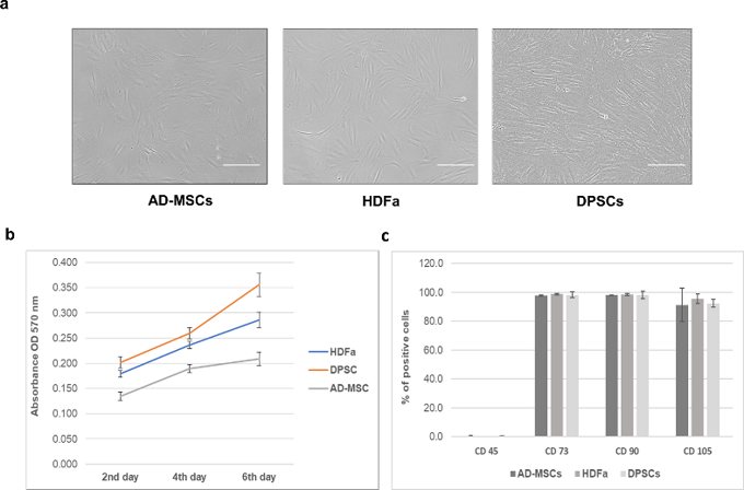
Mesenchymal stem cells (MSCs) are commonly used in regenerative medicine however the functional equivalency of MSCs from different tissue origins is not well established. Nováková et al. compared human dermal fibroblasts (HDFa) with dental pulp stem cells (DPSCs) and adipose-derived MSCs (AD-MSC) by proteomic and phenotypic studies in this study. All cell types stuck to plastic, had fibroblast-like shape and showed similar proliferation rates after six days (Fig. 1a, b). Flow cytometry revealed positivity for the mesenchymal stem cell markers CD73, CD90 and CD105 (>90%) and negativity for CD45 (<1%) (Fig. 1c).
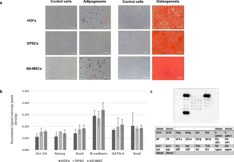
All the cell types showed trilineage differentiation potential except DPSCs which did not undergo adipogenesis (Fig. 2a). Proteome Profiler analysis of pluripotency markers showed similar Oct3/4, Sox2, Nanog, GATA-4, Snail and E-cadherin levels in all three sources with no significant changes (Fig. 2b, c). Untargeted proteomics did not detect these low-abundance proteins. These data show that HDFa, DPSCs and AD-MSCs have basic MSC properties and suggest their potential interchangeability in some therapeutic settings.
Ask a Question
Write your own review
Description: Origin: HumanDesease: Psoriasis (PS)Application: These cells can be used to study skin diseases such as wound healing, dermatitis, and other diseases that are expressed in fibroblast. In addition, ...
Description: Origin: HumanDesease: Arteriovenous Malformation (AVM)Application: These cells can be used to study skin diseases such as wound healing, dermatitis, and other diseases that are expressed in ...
Description: Origin: HumanDesease: GlioblastomaApplication: These cells can be used to study skin diseases such as wound healing, dermatitis, and other diseases that are expressed in fibroblast. In addition, ...
Description: Mesothelial cells play pivotal roles in ovarian cancer metastasis, peritoneal dialysis, surgical adhesions, inflammatory response, and metabolic disease. These specialized epithelial cells form a ...
Description: Origin: HumanDesease: Mucopolysaccharidoses (MPS)Application: These cells can be used to study skin diseases such as wound healing, dermatitis, and other diseases that are expressed in fibroblast. In ...
Description: Origin: HumanDesease: Systemic Lupus Erythematos (SLE)Application: These cells can be used to study skin diseases such as wound healing, dermatitis, and other diseases that are expressed in ...